Arthrodiastasis
1. General considerations
Bridging fixation with internal fixator
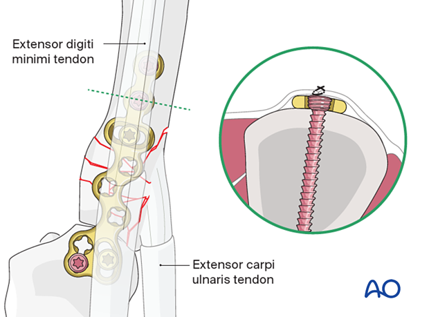
In severe comminution with a nonreconstructible joint surface, eg, a plate may not be applied to the articular block, a joint spanning plate may be used as a temporary internal fixator for arthrodiastasis, ie, allowing for the restoration of correct length and rotational alignment.
Reduction of the articular surface remains relevant.
A locking plate should be used to provide optimal stability and maintain length and alignment across the joint. It should be long enough to allow for two screws in each bone. Some special plates come with oblong holes that allow for adjustment of rotational and longitudinal alignment during plate application.
For example, to stabilize the 5th ray, a plate is applied dorsally with screws inserted into the hamate and the metacarpal diaphysis.
The plate is removed when the fracture has completely healed after about 4 months.
Associated dislocation of the 4th metacarpal
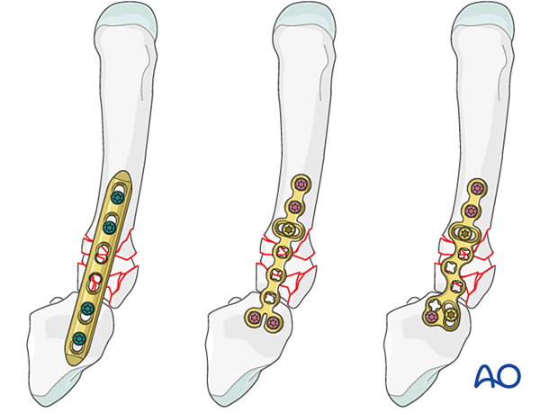
Fractures of the 5th metacarpal base may be associated with a carpometacarpal (CMC) joint dislocation of the 4th ray. In that case, the 4th metacarpal is reduced first and usually stabilized with K-wire transfixation.
Occasionally, there is a subluxation of the 3rd metacarpal or even the 2nd metacarpal, and any combination of additional fractures can be seen.
Once the dislocation of any other CMC joint has been stabilized, fixation of the fracture may proceed.
2. Approaches
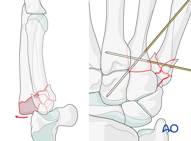
For this procedure, a dorsal approach to the carpometacarpal joints or a dorsal approach to the 5th carpometacarpal joint are normally used and enlarged as necessary to apply the plate.
3. Reduction
Restoration of length
Apply axial traction on the finger, either manually or with a finger trap.
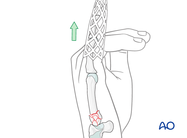
A small external fixator can be used, with K-wires inserted into the hamate and the distal metacarpal, preliminarily to align the ray and allow anatomical reduction of the joint surface.
The external fixator can be removed after finalizing plate application.
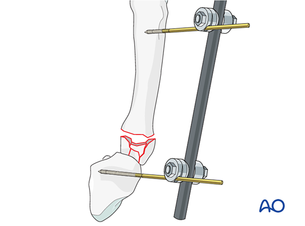
As an alternative to external fixation, a temporary K-wire may be inserted through the shaft and into the adjacent noncompromised metacarpal to maintain length and alignment. This K-wire can be removed after finalizing plate application.
Accompanying hamate fracture
If there is a shearing fracture of the hamate, reduce this fragment first and fix it with a lag screw.
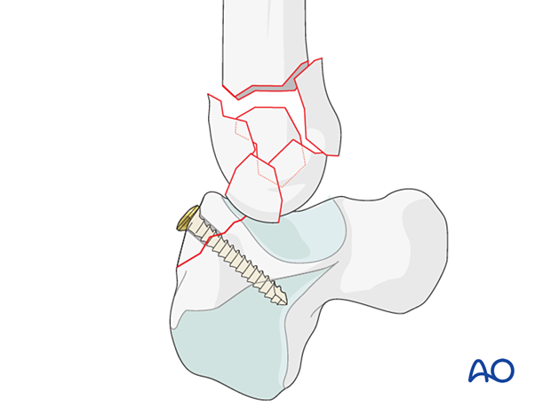
Reduction of fragments
Anatomical reduction and stabilization of the articular surface should be attempted although there is comminution.
A capsulotomy is needed to check the reduction of the articular fragments if the joint capsule is not already ruptured.
Use a dental, pick, periosteal elevator, or small K-wires to reduce the fragments. Insert small K-wires for preliminary fixation of these articular fragments. Occasionally, these K-wires are inserted percutaneously. Make sure not to injure the dorsal sensory branch of the ulnar nerve.
Check reduction using image intensification.
If the hamate is uninjured, its articular surface can be used as a template for restoring the articular surface of the metacarpal base.

Buttressing small fragments
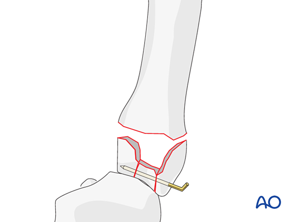
Additionally, a periarticular K-wires may be used to reposition and support impacted articular fragments.
The end of the K-wire may protrude through the skin, which facilitates removal after consolidation of the fracture.
Bone graft
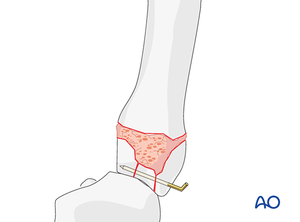
If conventional condylar plates are used, cancellous autograft, or very occasionally bone substitutes (eg, hydroxyapatite, tricalcium phosphate), will be necessary to support the articular fragments and fill the impaction void. Bone graft will enhance the healing process.
4. Checking alignment
Identifying malrotation
At this stage, it is advisable to check the alignment and rotational correction by moving the finger through a range of motion.
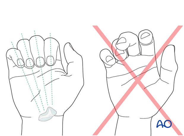
Rotational alignment can only be judged with flexed metacarpophalangeal (MCP) joints. The fingertips should all point to the scaphoid.
Malrotation may manifest by an overlap of the flexed finger over its neighbor. Subtle rotational malalignments can often be judged by a tilt of the leading edge of the fingernail when the fingers are viewed end-on.
If the patient is conscious and the regional anesthesia still allows active movement, the patient can be asked to extend and flex the finger.
Any malrotation is corrected by direct manipulation and later fixed. Flexing the MCP joints while preventing overlap of the fingers will reduce rotational displacement.
Using the tenodesis effect when under anesthesia
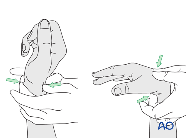
Under general anesthesia, the tenodesis effect is used, with the surgeon fully flexing the wrist to produce extension of the fingers and fully extending the wrist to cause flexion of the fingers.
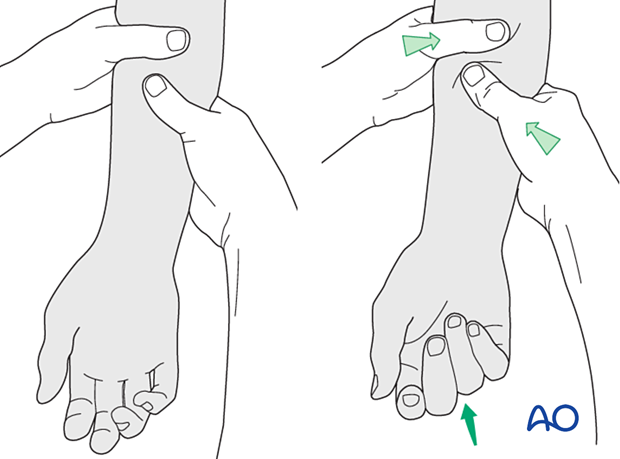
Alternatively, the surgeon can exert pressure against the muscle bellies of the proximal forearm to cause passive flexion of the fingers.
5. Plate application
Contour the plate exactly to fit the neutral position of the CMC joint, including any necessary twisting.
Confirm correct plate position with an image intensifier.
Insert a first locking head screw into the plate either in the carpal bone or the metacarpal diaphysis.
Check for correct rotational alignment of the ray clinically before inserting a screw into the other bone. Confirm again for correct rotational alignment.
Add at least another locking screw in each bone to finalize the fixation.
Be careful to ensure that the screws do not perforate the joint surface.
Cover the plate with periosteum to avoid adhesion between the tendon and the implant leading to limited finger movement.
