Plate fixation
1. Principles
Restoring normal anatomical contour is crucial to avoid long-term complications such as loss of reduction, facial asymmetry and general dysfunction.
2. Approach
Patient positioning
For this procedure, one of the following two patient positions are used:
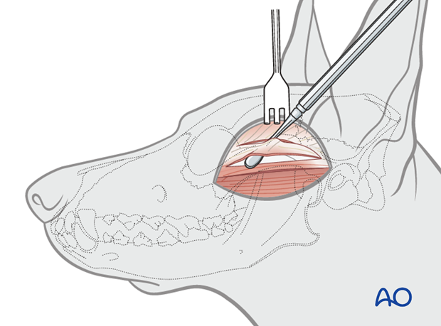
The lateral extraoral approach to the zygomatic arch allows for the best exposure.
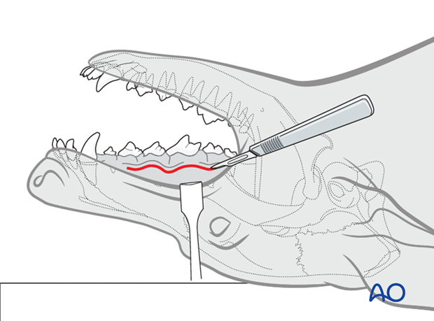
If only the rostral part of the zygomatic arch is fractured, an intraoral approach may be attempted.
3. Irrigation of the fracture
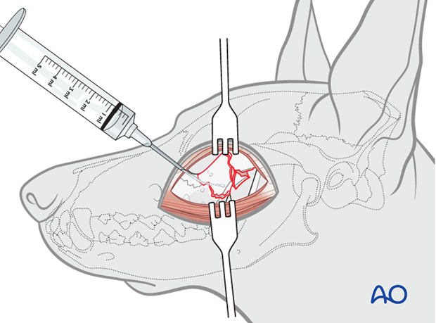
Irrigation of the fractured area is advised to remove any foreign material and debris.
4. Reduction
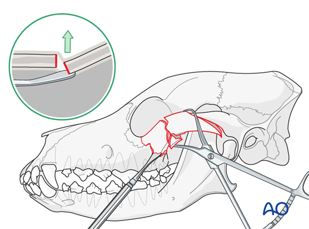
The fracture area is inspected for non-vital structures. If non-vital structures are identified, they should be removed and discarded.
The dislocated segment is elevated or distracted using one or two periosteal elevators and/or bone-holding forceps until the appropriate anatomic alignment is reached.
5. Fixation
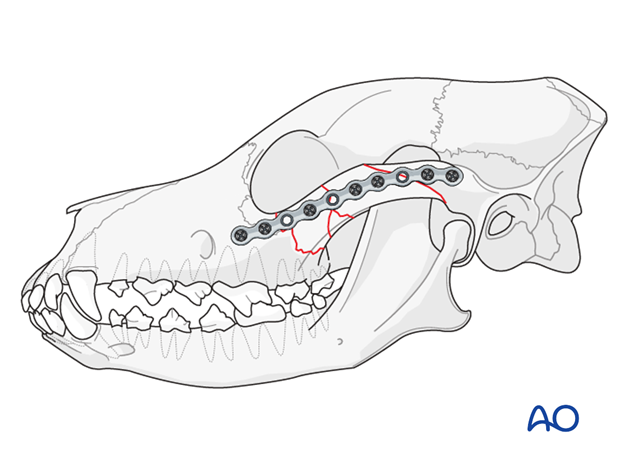
An appropriate size, low-profile, titanium 2mm non-locking miniplate is contoured using specially designed miniplate bending pliers, and adapted to the desired anatomical site. This allows for accurate repositioning and reconstruction of the zygomatic arch. The length of the plate should allow for at least two screws on each side of the fracture. Three screws per fracture fragment (six cortices) are ideal.
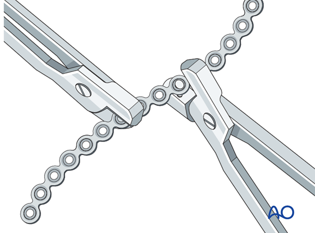
Plate application
The plate is applied spanning the fracture margins. More than one plate is rarely required.
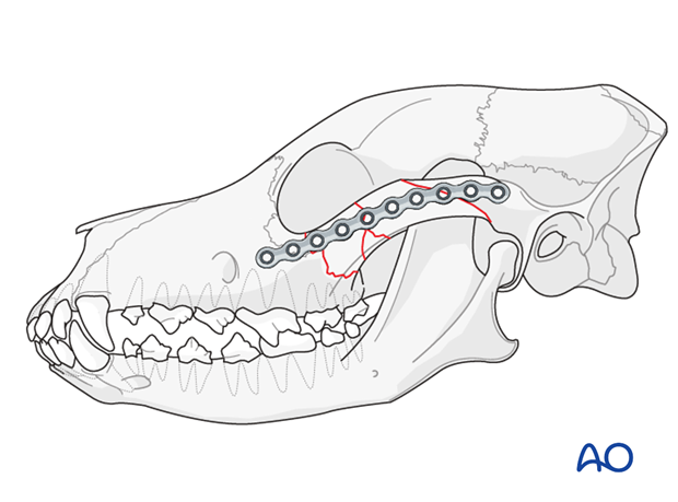
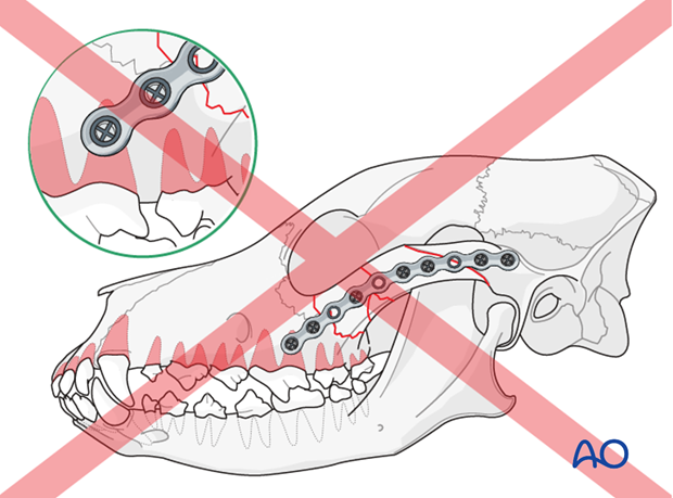
Note: Care should be taken with placement of the rostral screws to avoid the roots of the maxillary molar and fourth premolar teeth.
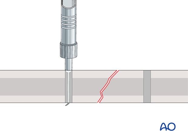
The length of the screws is determined by using CT measurements of the bone thickness and a depth gauge.
The plate is secured to the bone with at least two non-locking, self-tapping titanium screws in each segment of the fracture.
6. Closure
The soft tissues are closed routinely in two layers. A Stent bandage is secured in place for 72 hours in order to minimize postoperative swelling and emphysema.
7. Case example
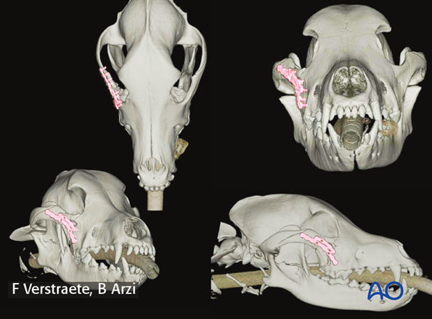
A 1.5-year-old FS Labrador Retriever with a minimally comminuted and depressed zygomatic arch fracture, from being kicked by a horse.
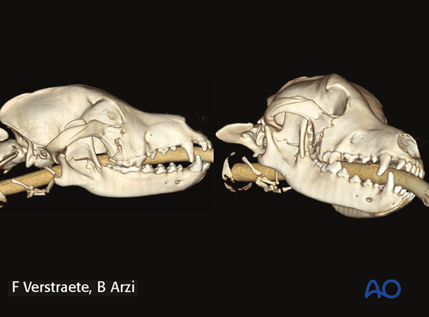
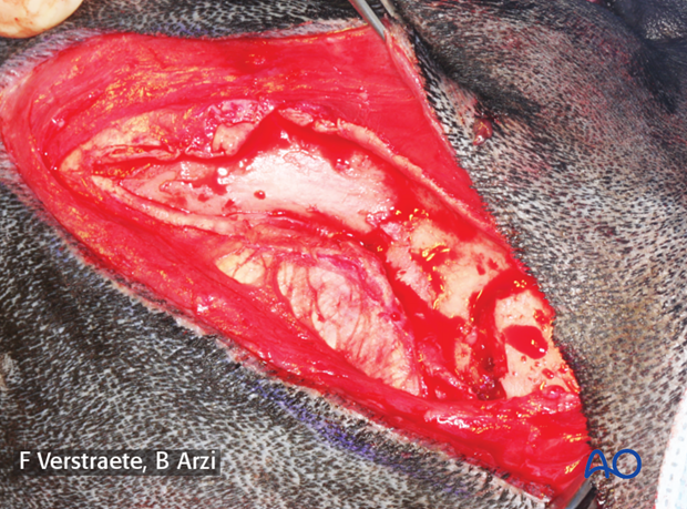
Lateral approach to the zygomatic arch was performed to expose and reduce the fracture.
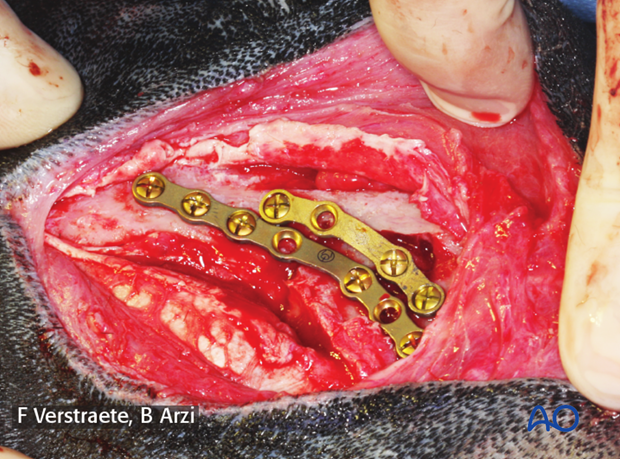
Two 2.0mm non-locking titanium miniplates were used to restore continuity and the attachment of the orbital ligament to the frontal process of the zygomatic bone.
Postoperative CTs at three months showing bone healing and remodelling.
8. Aftercare
The dog should be kept on soft food for one or two weeks after surgery.
Intraoperative intravenous administration and then oral antibiotics for one to two weeks is recommended. Antibiotics used should have an excellent oral cavity penetration and spectrum. Example: ampicillin at 20 mg/kg intravenous antibiotics followed by oral antibiotics amoxicillin/clavulanic acid at 15-20 mg/kg orally twice daily.
Pain medication, such as opioids and non-steroidal anti-inflammatory medications, are used for 7-14 days.
If the fracture involved the oral cavity, oral rinse with 0.05-0.12% chlorhexidine gluconate solution is recommended (not for maxillomandibular fixation).
Recheck is recommended in 10-14 days and at three and six months post-surgery.
If there are any signs of complications (i.e. purulent nasal discharge), CT and rhinoscopy are indicated.
Implant removal
Typically, titanium miniplates and screws are osseointegrated. If there is no implant failure or infection, there is no need for implant removal.


