Intraoral approach to the maxilla
1. Indications
The intraoral approach is used to expose and reduce fractures of the caudal or rostral maxilla if they are poorly accessible by an extraoral approach. This approach allows for better visualization of fractures near the dentoalveolar structures.
2. Mucosa incision
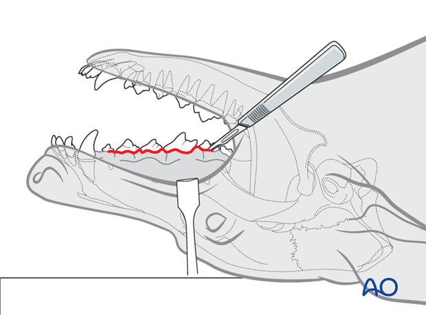
For better exposure, the dog should be placed in dorsal recumbency as demonstrated in this image.
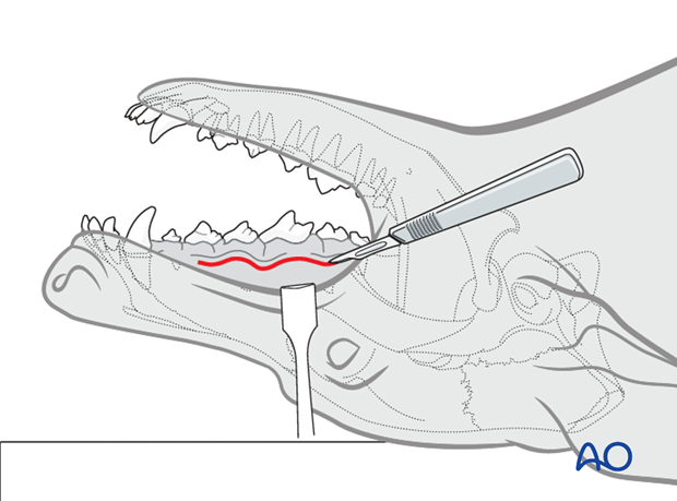
Using a N°15 scalpel blade, the incision is placed 3-5 mm away from the mucogingival junction.
The incision extends as far caudally and rostrally as needed to provide appropriate exposure for reduction and placement of internal fixation. A caudal incision will encounter the facial vein as it joins the deep facial vein near the levator nasolabialis muscle.
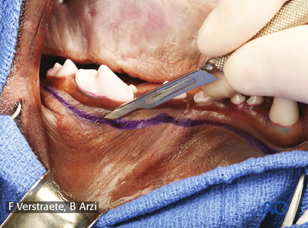
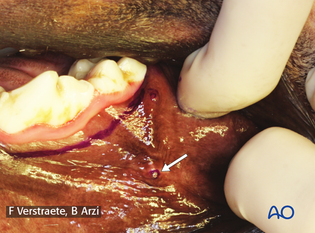
Care should be taken to avoid damage to the parotid and zygomatic salivary papillae.
Alternatively, a sulcular incision is made followed by elevation of the gingiva and the mucosa with a periosteal elevator (as performed for surgical extractions).
3. Exposure
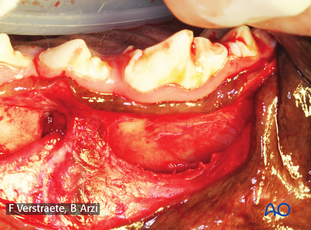
Subperiosteal elevation is performed using a periosteal elevator to elevate the mucosa, submucosa, and facial muscles. Care should be taken to identify and isolate the infraorbital neurovascular bundle. Damage to these muscles should be avoided, as it can compromise the blood supply to the fractured area and may cause loss of sensation.
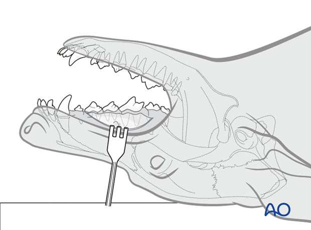
Pearl: Once the bundle and the foramen are identified, the periosteum should be elevated completely around the foramen. This allows for better mobility and manipulation of the infraorbital bundle.
4. Closure
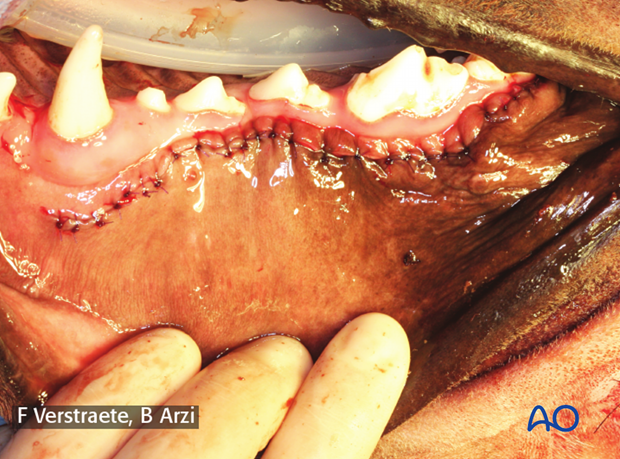
Closure can be done in one or two layers using 4.0 poliglecaprone 25 (or other monofilament absorbable suture) in a simple-interrupted pattern.
