Patient assessment
1. General considerations
High-energy trauma may be associated with greater fragmentation and soft-tissue damage, including a greater risk for neurovascular injuries.
Low-energy trauma in poor-quality bone of elderly patients may be associated with greater fragmentation of the articular block.
The fracture may be caused by axial loading or a direct impact on the elbow joint. Often, the patient may not recall the exact mechanism during the trauma.
2. Case history
It is essential to obtain a comprehensive history. This should consider the mechanism of the injury and additional injuries and hand dominance, activity level, personal needs, and any comorbidities.
3. Clinical examination
Examine the elbow, forearm, and hand for:
- Deformity
- Open wound
- Swelling
- Bruising
- Neurologic or vascular deficit (especially ulnar and radial nerves)
Evaluation and documentation of the radial, posterior interosseous, ulnar, and median nerves for a “primary nerve injury” is essential before any reduction maneuver.
The presence of a radial pulse at the wrist does not necessarily imply that the brachial artery is not injured: collateral perfusion can bypass a damaged brachial artery. If there is any doubt, use a Doppler ultrasound scan to determine the integrity of the brachial artery.
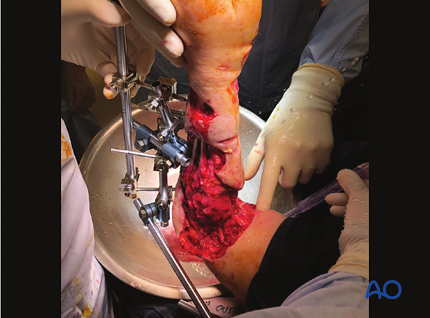
An adjacent wound implies an open fracture, which needs urgent surgical debridement.
4. Open fractures
Open fractures may occur due to high energy mechanisms or in the presence of a fragile soft-tissue envelope around the elbow, such as in elderly patients.
Be aware of a higher incidence of neurovascular injuries and associated bony or other system polytrauma.
Neurovascular injuries need urgent assessment and management. The treatment options, including operative approach, type of fracture fixation, and surgical management, are determined by the neurovascular state. In polytraumatized patients, the management of the elbow fracture is subordinate to the overall resuscitation of the patient and may be delayed. In this situation, temporary fixation of the limb in anatomic alignment (splinting or external fixation) may help protect the soft tissues until definitive treatment is possible. Frequent and regular observations of neurovascular status are mandatory.
The gold-standard treatment is early debridement, intravenous antibiotics, bony stabilization, and soft-tissue management. A combined orthoplastics approach can be helpful.
A staged approach may be optimal. This includes initial soft-tissue and bony debridement, temporary skeletal stabilization (external fixation) followed by serial debridements, and definitive fixation when soft-tissue coverage is possible. Vacuum-assisted wound management may be helpful.
Restoration of perfusion of the forearm and hand should take priority: vascular repair or bypass should be performed primarily after emergency stabilization of the elbow region using external fixation.
Damaged nerves should be tagged for later identification and repair during subsequent procedures.
5. Radiological examination
General considerations
Take x-ray images in two perpendicular planes showing the elbow, distal humerus, and the entire forearm (including the wrist). This allows for assessment of longitudinal and rotational alignment and associated injuries of the wrist.
If emergency x-ray imaging is insufficient for diagnostic purposes, a planned examination under anesthetic with x-ray views under traction is very helpful.
Decision-making, including preoperative planning, is optimized by 2-D and 3-D CT scanning.
Fracture assessment
X-ray imagingSometimes the appearance of a fracture pattern is more complex, but it can be difficult to understand it on standard x-rays, particularly if a cast obscures the view.
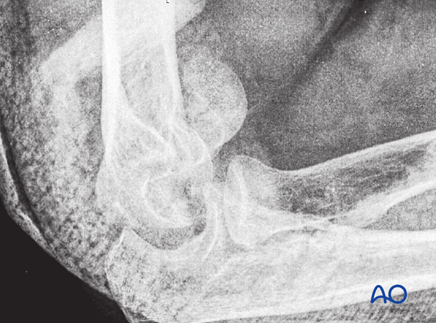
Computed tomography, with 3-D reconstruction particularly, is very useful for understanding apparently simple fractures that become more complex.
Apparently nonarticular medial or lateral condylar fractures
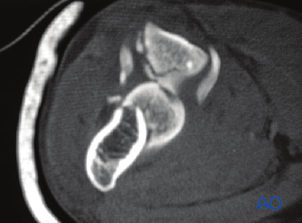
Take care when assessing any apparently nonarticular medial or lateral condylar fracture, as these are unusual. Preoperative imaging, including computed tomography, can be used to identify associated articular fractures.
This image shows a 2-D CT scan of a medial sagittal, simple transtrochlear fracture.
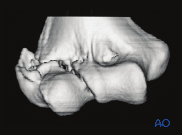
This image shows a 3-D CT reconstruction of a lateral sagittal, simple transtrochlear fracture. Although the major fragments are clearly seen, the intrafocal comminution is obscured by virtue of the method of image rendering.
Apparently simple capitellar fractures
Take care when assessing an apparently simple capitellar fracture. These are often complex. The majority extends into the lateral trochlea.
Many fractures also involve a fracture of the lateral epicondyle and impaction of the posterior aspect of the lateral column or the trochlea.
Computed tomography with 3-D reconstruction is necessary to identify the fracture characteristics and facilitates planning.
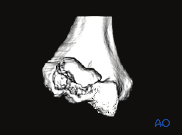
Trochlear fractures
Trochlear fractures are difficult to see on radiographs. Computed tomography—with 3-D reconstruction—is particularly useful for understanding fracture anatomy.
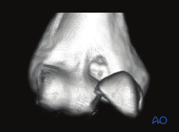
There is often complex articular comminution.
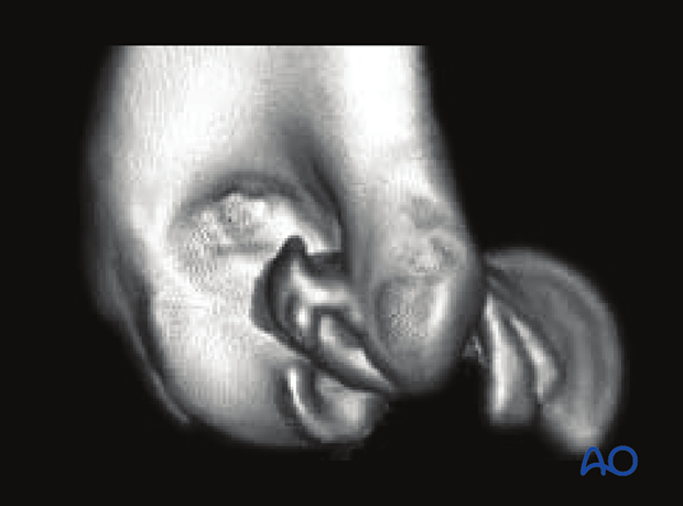
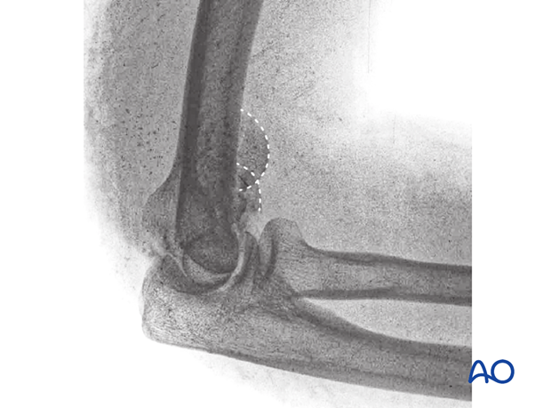
Coronal fractures involving capitellum and trochlea
The “double arc” sign indicates the involvement of the trochlea in a coronal fracture.
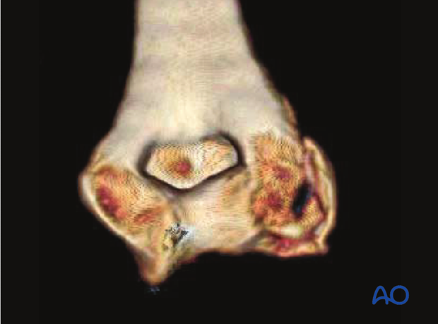
This case shows a complex coronal fracture of capitellum and trochlea, involving a lateral epicondylar fracture, posterior impaction, and medial fracture extension.
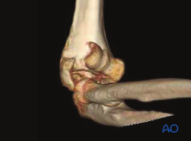
Here, it is apparent that the lateral epicondyle is fractured.
The posterior aspect of the lateral column is also fractured.
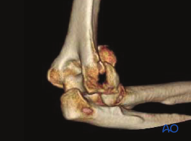
In this image, it is apparent that the articular fragments may not correctly align, suggesting impaction of the posterior trochlea and particularly the lateral column.
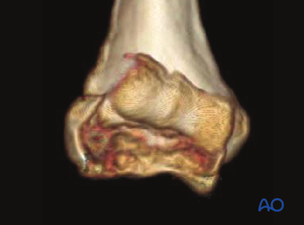
An end-on view shows the complexity, particularly on the lateral side.
Also, one can see a fracture—albeit very subtle—between the medial epicondyle and the medial trochlea.
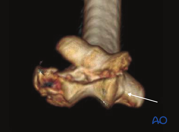
Here, the medial-side fracture can be seen and what appears to be impaction (narrowing of the olecranon fossa). These injuries are very subtle but were confirmed during operative exposure.
6. Further investigation
If there is concern about a neurological or vascular injury, see the corresponding additional material


