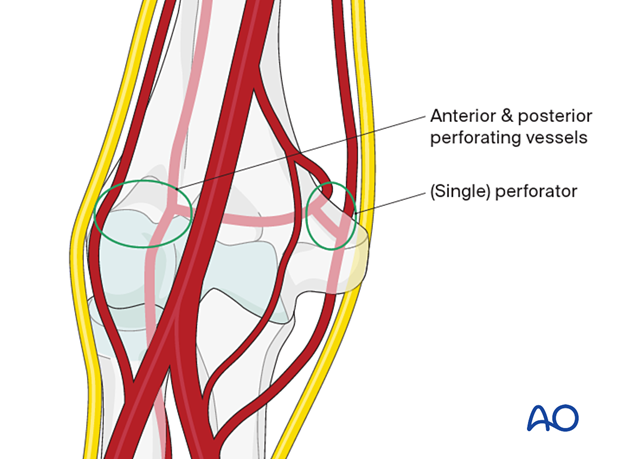
Vascularization of the distal humerus
1. Introduction
The vascular supply of the elbow follows the same overall pattern of perfusion of all epimetaphyseal regions:
- Each collateral vessel contributes to a transverse anastomotic system proximal and distal to the articular segment.
- The anastomotic systems contribute endarteries to the bone.
- The endarteries anastomose within the bone in a constant manner at critical zones of bone perfusion.
The preservation of perfusion is critical for the healing process and survival of the distal humeral articular segments.
The surgical approaches all potentially place the vascular supply at the elbow at further risk.
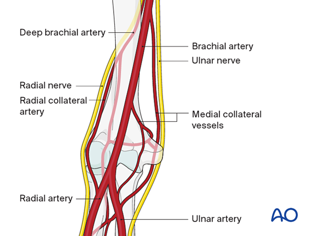
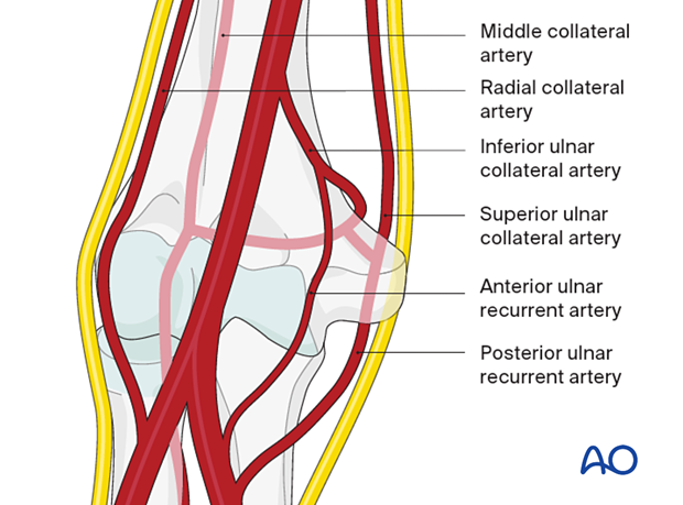
2. Medial collateral vessels
There are longitudinal collateral vessels on the medial side:
- The superior vessel passes with the ulnar nerve posterior to the medial epicondyle.
- The inferior vessel passes anteriorly to the trochlea.
Both anastomose with the ulnar artery distal to the elbow.
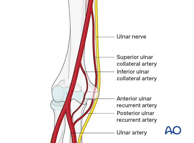
3. Lateral collateral vessels
The lateral collateral vessels are based on the deep brachial artery:
- The deep brachial artery passes in front of the lateral epicondyle with the radial nerve. It anastomoses with the radial artery distal to the elbow.
- The radial collateral artery is a branch of the deep brachial artery and passes posterior to the lateral epicondyle. It anastomoses with the interosseous artery distal to the elbow. This anastomosis is variable.
Principle: There are longitudinal vessels from the brachial artery, two on each side. Each pair of vessels comprises one anterior and one posterior on each side of the distal humerus.
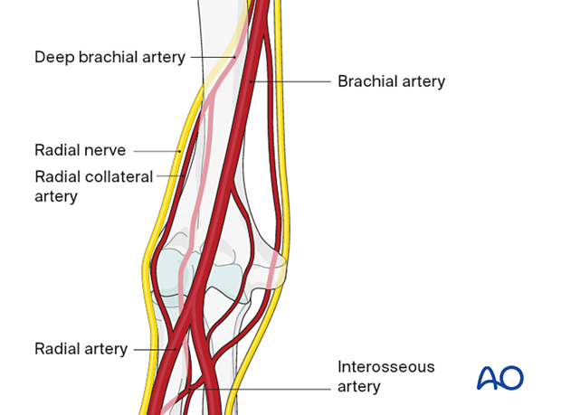
4. Critical anastomosis
There is a transverse anastomosis at the level of the olecranon fossa between the two posterior collateral vessels (the superior ulnar collateral and deep brachial arteries):
- This also communicates with the anteromedial (inferior ulnar collateral artery) system.
- In all supracondylar fractures, this anastomosis is easily interrupted.
5. Perfusion of the articular block
- Perfusion of the medial side of the distal humerus is through a usually single perforator from the medial side of the critical anastomosis.
- Perfusion of the lateral side of the distal humerus is through multiple perforators anteriorly and posteriorly from the robust anterior and posterior collateral vessels.
Principle: The perfusion of the lateral part of the distal humeral articular block is more robust than the perfusion of the medial part.
- The intraosseous anastomosis between the perforating vessels occurs in the lateral part of the trochlea.
- Fractures that separate the lateral part of the articular block from the medial part may result in ischemia of the most lateral extent of the medial part of the articular block.
- Complex fractures of the articular block that isolate a segment of the trochlea from both medial and lateral parts often result in ischemia of that segment.
Principle: Preservation of collateral blood supply to the distal humerus by respecting the soft-tissue attachments to the condyles is important to the perfusion of the articular block.
