Clinical and radiographic examination
1. Etiology
Femoral fractures are most commonly diagnosed in foals. They rarely occur in adults, and when they do occur the bone is damaged beyond repair.
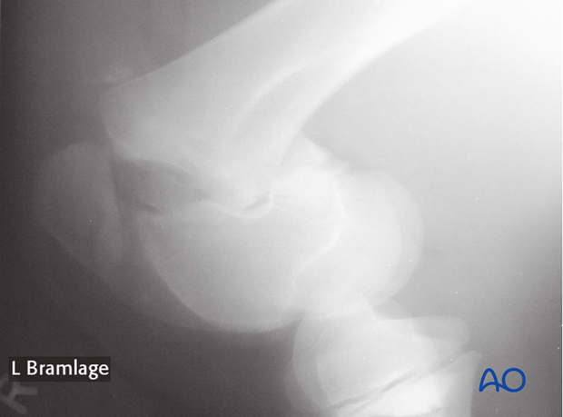
Femur fractures most commonly occur when the foal falls on the affected limb. If the foal falls with the limb across his body, in the adducted position, a spiral mid-shaft fracture most commonly results. If the foals foot is trapped in a fence or under an obstacle and the animal subsequently falls with the limb hyperextended, a Salter-Harris type II distal femoral fracture will result.

2. Fracture types overview
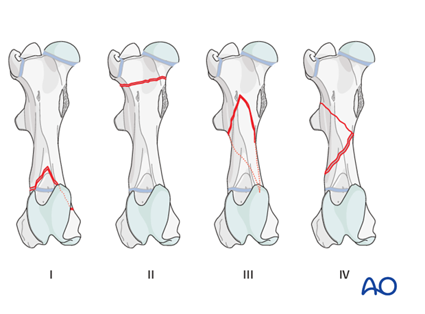
Four common fracture types occur in the horse’s femur:
- Salter Harris type II distal femoral physeal fractures.
- Proximal subtrochanteric fractures
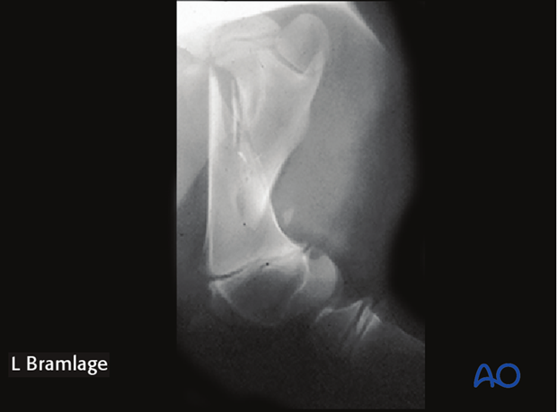
- Spiraling mid-diaphyseal fracture
- Comminuted fractures
Salter Harris type I proximal femoral physeal fractures are very rare and they are rarely treated. Therefore these fractures will not be addressed in this module.
In this module, treatment options will be described for Salter Harris type II fractures and spiraling mid-diaphyseal fractures.

3. Clinical signs
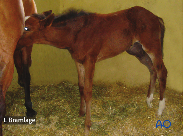
Clinical signs are non weight bearing lameness with mild to severe swelling of the thigh region of the horse.

4. Imaging
Radiographs of the horse’s femur are difficult to take. The distal femur can be radiographed from lateral to medial in the standing horse. Radiographs of the complete femur require lateral recumbency with the affected limb down and medial to lateral direction of the x-ray beam.
Ultrasonography can be used to confirm the presence of a fracture but is not useful to determine the fracture configuration.

5. Definition
Salter Harris type I distal femoral physeal fractures occasionally occur, but they are normally stable and do not require surgical treatment, just exercise restriction.


