Lateral approach to the equine femur
1. Lateral approach
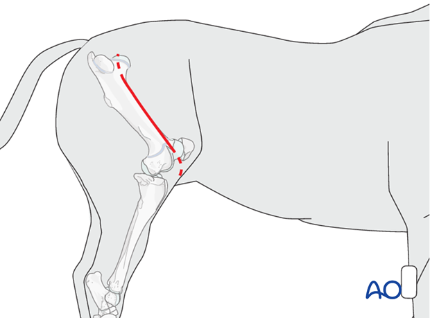
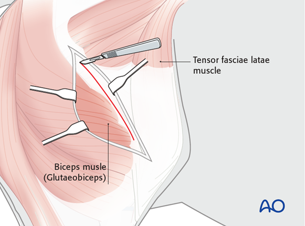
The incision is made from the lateral aspect of the greater trochanter to the lateral aspect of the patella following the junction of the biceps femoris and the vastus lateralis muscles.
The incision can be extended cranially at the proximal extent for access to the femoral neck or can be extended caudally at the distal end along the path of the lateral patellar tendon for access to the stifle joint and distal femoral condyles.

2. Deep dissection
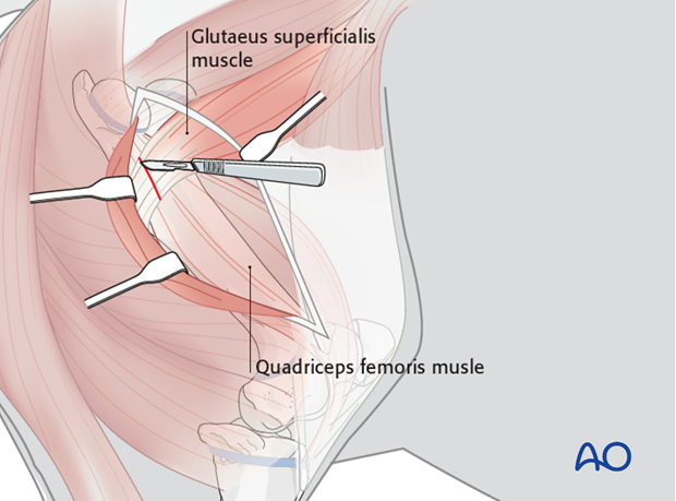
The vastus lateralis and biceps femoris muscles are separated. The superficial gluteal tendon must be transected from its attachment to the third trochanter.
Enough tendon needs to be left attached to the trochanter to allow reconstruction of the transected tendon at closure.


3. Closure
Antibiotic containing implants (PMMA beads or collagen sponges) are placed alongside the plates. Suction drainage is always used in femoral fractures because of the large muscle mass surrounding the femur and its involvement in the trauma of femoral fractures.
The injured muscles produce considerable hemorrhage and serum, which must be evacuated from the fracture site to minimize the chances of infection.
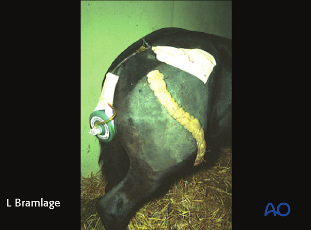
The fascia overlying the muscles of the thigh are closed as the first layer. The subcutaneous tissue and the skin are closed sequentially. A stent bandage is sutured over the incision.

The suction drainage catheter can be seen exiting the proximal aspect of the fracture cavity above the incision. The suction reservoir is attached to the horse’s tail.
A stent bandage has been sutured over the incision for recovery.
The suction drainage is removed when the production of fluid begins to decline at 3-4 days postoperatively. The stent bandage is normally removed at 5-7 days postoperatively.


