Plate fixation
1. Principles
Restoration of dental occlusion and normal anatomy is crucial to avoid long term complications such as malocclusion, malunion, nonunion, facial asymmetry and loss of function.
2. Approach
Pharyngeal intubation is needed to assess and maintain occlusion throughout and following the surgery.
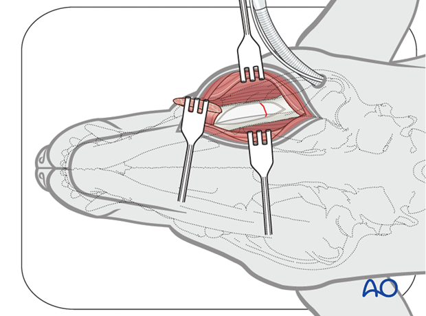
With the patient in dorsal recumbency, a ventral approach to the ramus of the mandible or mandibles is performed. The incision should expose enough intact bone to place at least two screws on both sides of the fracture.
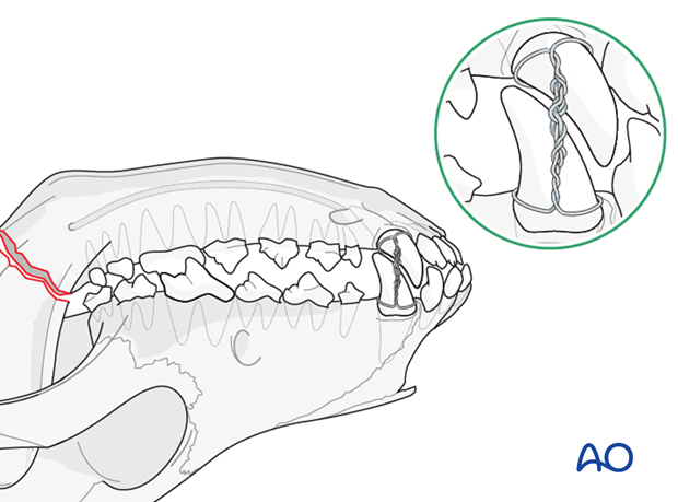
The mandible and maxilla should be wired together to lock the mouth into normal dental occlusion. This will help reduce the fracture(s) and ensure proper postoperative alignment of the fracture(s).
The technique uses 26 or 28 gauge wire. The wires are looped around all four canine teeth and the maxilla and mandibular wires on each side are twisted together.
3. Reduction
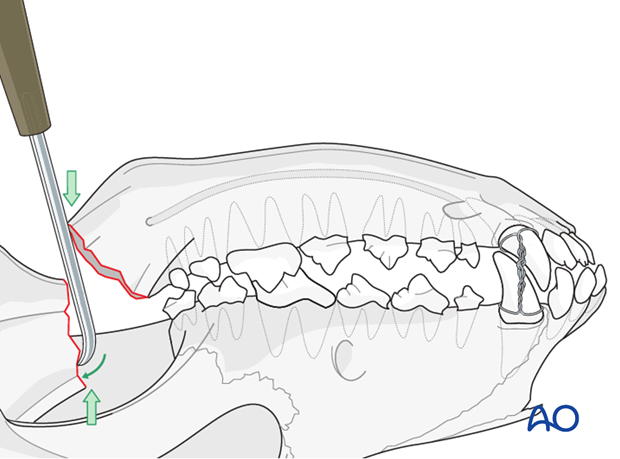
Fracture reduction is accomplished with 1-2 periosteal elevators placed at the fracture site to lever the caudal ramus fragment into reduction to restore the normal anatomy. Bone holding forceps and digital reduction can be used.
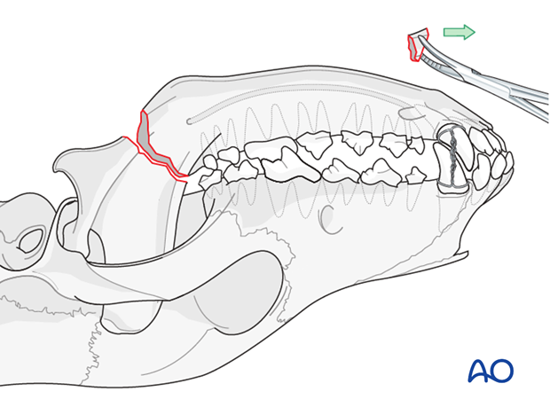
Small loose fragments with no soft tissue attachment should be removed to avoid possible sequestrum formation.
4. Fixation
Plate selection and contouring
For a medium or large breed dog, a single 2.4/3mm titanium locking mandibular reconstruction plate is used. For small breed dogs and cats, a 2.0mm titanium locking plate or mandibular reconstruction plate is used.
Note: If the caudal ramus fragment is too small for placement of two locking screws, a minimally invasive fracture fixation instead of bone plating should be used.
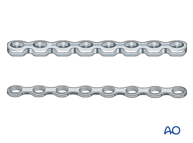
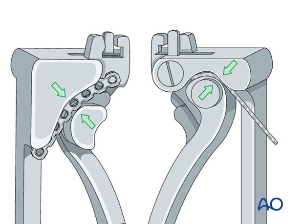
The mandibular reconstruction plate is anatomically contoured in 3 dimensions using the appropriate bending pliers.
The length of the plate should allow for 2-3 bicortical locking screws in each fracture segment.
Plate application
The plate is applied to span the fracture site and screw placement must avoid the mandibular foramen. The plate is placed ventral to the mandibular canal to avoid the mandibular foramen. A single plate is sufficient to stabilize the mandible.
Note: In small dogs and cats, due to limited bone stock, the screws often penetrate the mandibular canal in the rostral fragment. The inferior alveolar neurovascular bundle is already damaged from the fracture, therefore penetrating the mandibular canal is acceptable.
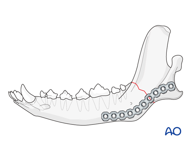
Plate fixation
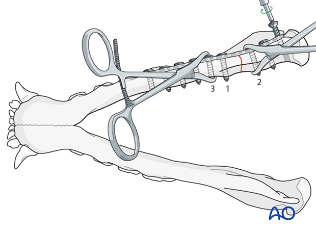
The plate is held in place using bone holding forceps. A locking drill guide is screwed into the plate to allow precise drilling of the hole.
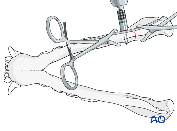
The rostral screw closest to the fracture site should be inserted first alternating as illustrated until all screws are inserted.
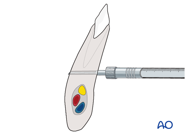
The screw length is determined by a depth gauge measurement. Preoperative CT measurements can be used to estimate the screw length for planning purposes.
5. Closure
The soft tissues are closed routinely in 3 layers.
6. Fractures of the ramus of the mandible including the coronoid process
A fracture involving both the coronoid process and the ramus of the mandible may be treated with:
- A single 2.4mm locking reconstruction plate (for medium to large breed dogs) or 2.0 locking plate (for small dogs and cats) to stabilize the ramus fracture
- Maxillomandibular fixation (MMF) if it's a simple ramus fracture in a young animal, with minimal displacement. Rigid fixation with the bone plate is preferred.
7. Case example
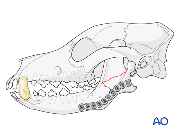
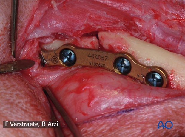
Simple mandibular fracture of the ramus in a small dog repaired using a 2.0mm locking titanium plate and locking screws.
8. Aftercare
Multimodal analgesia is recommended. Non-steroidal medication for 10-14 days and opioids for the first 5 days post-surgery as needed. Antibiotic therapy is prescribed for a period of 10-14 days following surgery.
Soft food should be fed for the first 14 days, followed by a gradual return to eating kibble over 2 weeks. Rough play (i.e., tug of war) should be avoided for the first 3 months after surgery.
Suture removal is performed 10-14 days after surgery.
A radiographic recheck is performed every 6 weeks until the fracture is healed. The plate is not removed after the fracture is healed.


