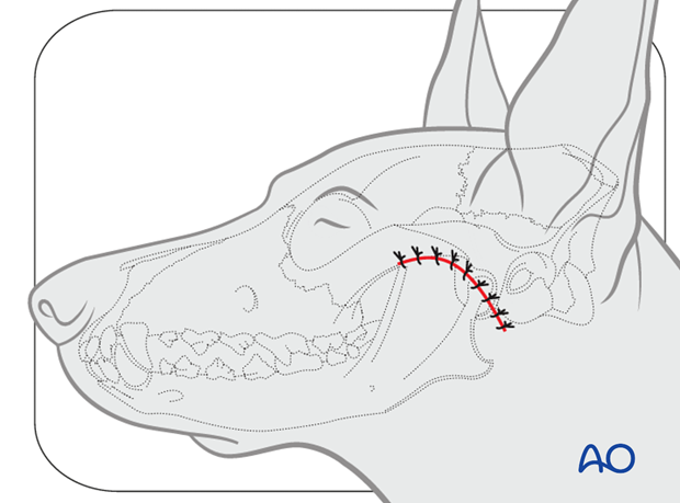
Lateral approach to the TMJ
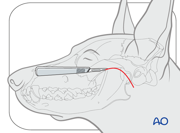
1. Skin incision
The lateral approach to the TMJ is performed with the patient in lateral recumbency.
The skin incision follows the ventral border of the zygomatic arch and is angled to cross the TMJ caudally.
2. Exposure
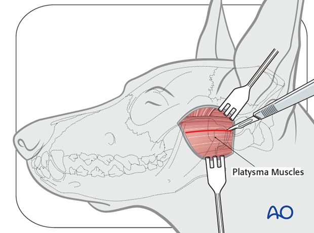
The platysma muscle is incised.
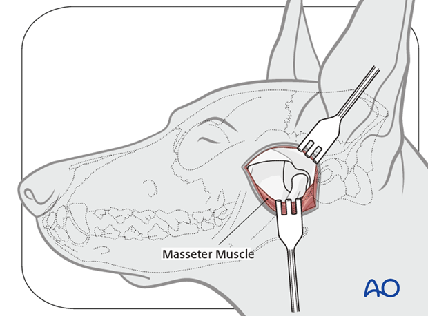
The origin of the masseter muscle is incised.
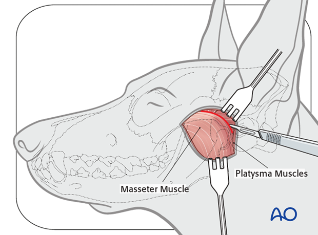
The masseter muscle is retracted and the TMJ is identified.
3. Closure
Closure is done in three layers. The first layer is the elevated muscles. The platysma and subcutaneous tissue is the second layer, followed by the skin.
Closure of the first two layers is done with absorbable sutures such as 4.0 910 or poliglecaprone 25. The skin is closed with monofilament nonabsorbable sutures, in a simple-interrupted fashion.
