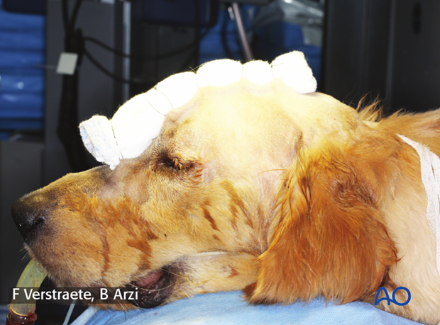
Dorsal midline approach
1. Indications
The dorsal midline approach is indicated for fractures of the frontal, nasal, and maxillary bones. It provides an excellent view of the fracture area, allows fracture reduction, and placement of implants.
2. Skin incision
The length of the incision is determined by the fracture location. The use of electrocautery should be avoided.
An incision is made using a scalpel blade extending caudally and rostrally as needed.
Fractures of the maxillary and nasal bones require an incision from the incisive bone to the level of the frontal bone.
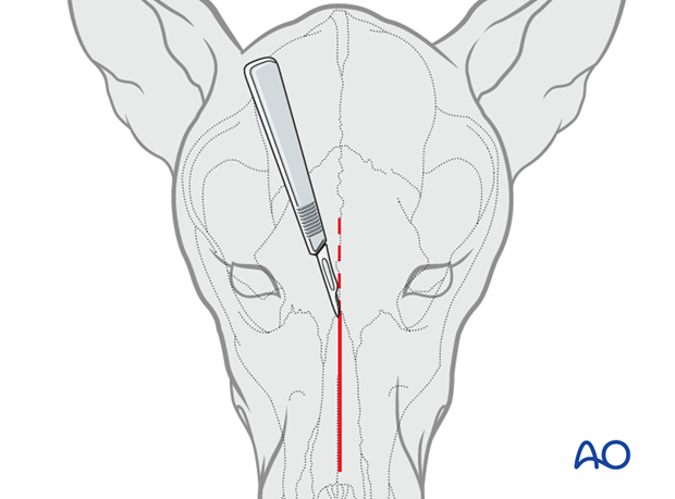
For frontal sinus fractures, the incision begins between the orbits and extends caudally to the rostral end of the sagittal crest.
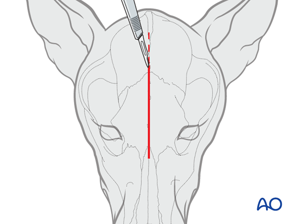
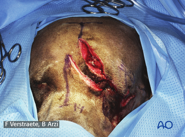
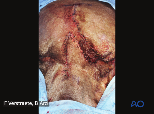
A standard dorsal midline approach to the fracture should always be used even when there are wounds in the area. Ideally, avoid going directly through the traumatic wound to prevent unnecessary trauma to the injured area. If the wounds are at the location of the standard approach, then going through the wounds may be necessary.
Note how the traumatic wound is avoided in the image.
The exposure should be large enough for proper inspection and reduction of the fracture. The wound must be debrided gently, and vital tissues preserved.
3. Exposure
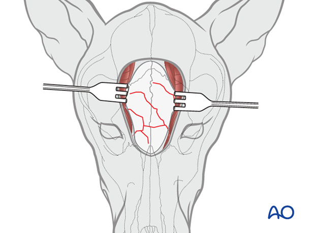
The superficial muscles (frontalis and intercutularis) are incised and reflected. The periosteum is elevated with a periosteal elevator. Excessive force should be avoided as the bone is very thin and can be easily perforated.
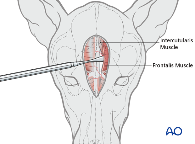
The tissues are then retracted to allow visualization of the entire fracture area.
4. Closure
Closure is done in two or three layers: periosteum, superficial muscle, and connective tissues in one or two layers; the skin in the second/third layer.
A Stent bandage may be used to prevent postoperative emphysema and to reduce swelling.
