Posterior midline approach to the sacrum (S1–S5)
1. General considerations
The exposure for resection of primary tumors may be wider than in a trauma approach. Moreover, more lateral extension may be required. In the case of previous intralesional surgery, wound excision is advised to remove the scar as it may have been contaminated by the tumor during the first surgery. Lastly, some patients will need preoperative radiation therapy as part of their treatment plan, which could render the approach more difficult and more prone to postoperative complications. Meticulous surgical technique in the exposure and closure is warranted.
2. Skin incision
The location and length of the skin incision varies according to the pathology being treated.
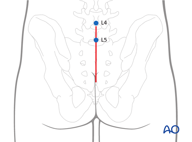
Use a midline incision to expose both sides of the sacrum.
3. Transection of the muscle aponeurosis
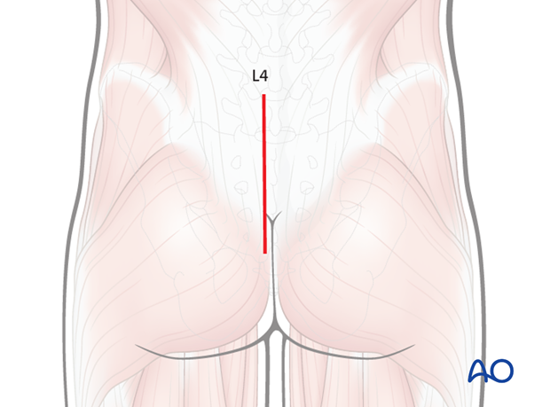
Extension of the exposure to the lumbar spine may be required on a case-by-case basis. If required, incise the lumbosacral fascia parallel to the spinous processes L4 and L5.
4. Dissection
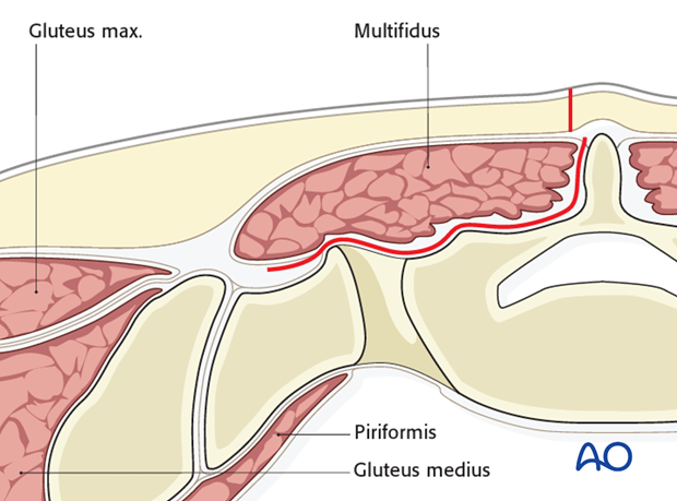
Subperiosteal elevation of the Iumbosacral musculature exposes the posterior aspect of the sacrum.
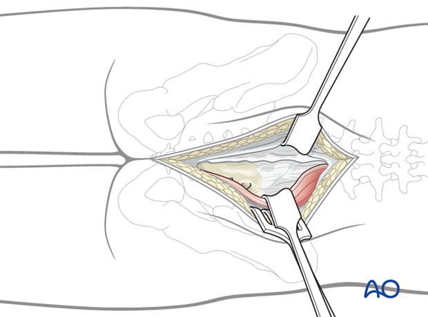
This is typically done by incising the multifidus insertion medially and elevating it laterally.
If a more extensile exposure of the lateral sacral region (ala) is necessary, elevate the complete muscle mass by dissecting its lateral attachment to the posterior iliac crest.
5. Wound closure
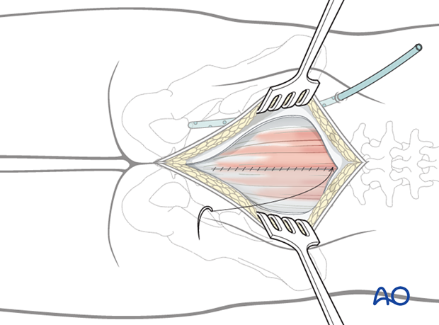
Initiate the closure of the wound by a gentle repositioning of the erector spinae muscles and reattachment of the superior origin of the gluteus maximus.
For patients undergoing revision surgery and/or with history of radiation, plastic surgery should perform the soft tissue reconstruction to decrease the risk of wound complications.
Drains are usually inserted via a separate stab incision.
Close the subcutaneous tissues and skin in layers.
