Prone for T1-S1
1. Positioning for midline approach (posterior procedures)
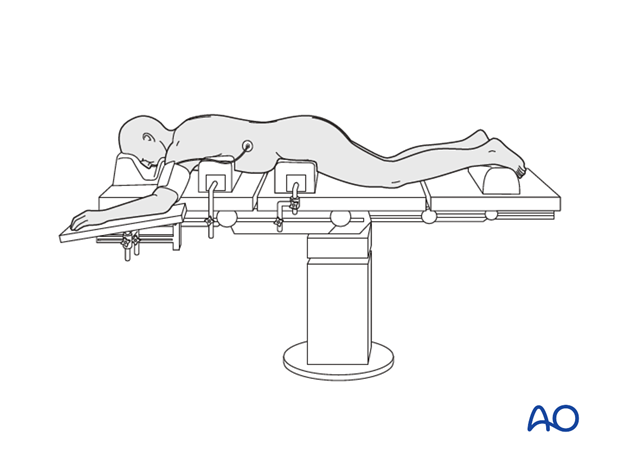
The patient is positioned onto a radiolucent table prone on two horizontally placed padded bolsters (one at the level of sternum and another one at the level of anterior iliac spine) or a frame.
- The abdomen should hang free to avoid increased intraabdominal pressure to prevent excessive bleeding
- Adequate padding needs to be provided to elbows and knees to avoid pressure sores
- The head is rested either in a padded head holder or a Mayfield rest to avoid pressure on the eyes.
Make sure that there are adequate personnel to receive and turn the patient from supine to prone position on the operating table.

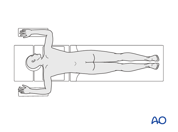
For fixations that start below T8, the arms can be abducted and should be resting comfortably at 90° position of the shoulder and elbow.

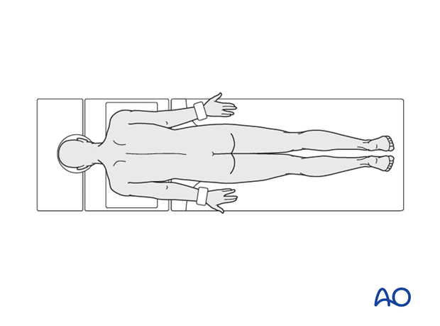
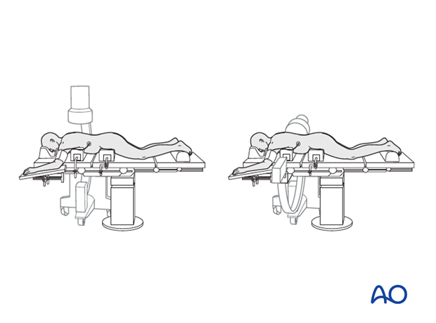
For fixations that extend above T8, the arms are adducted at the shoulder and extended at the elbow and strapped to the sides of the body. This helps in the hassle free use of image intensifier for the visualization of thoracic vertebra.

2. Anesthesia
General anesthesia with endotracheal intubation is required.
In patients with severe spinal cord compression, it is essential to avoid hypotensive anesthesia and the mean arterial blood pressure should be maintained above 80 mmHg.
3. Preoperative antibiotics
Antibiotics should be administered well prior to the incision and also at intervals during the procedure or when the blood loss exceeds 2 liters.
A cephalosporin antibiotic with good Gram-positive coverage is generally recommended.
4. Spinal cord monitoring
Spinal cord monitoring is optional.
5. Fluoroscopy/x-ray control
The incision can be planned based on the lateral fluoroscopic view.


