Medial approach to the proximal tibia
1. Patient positioning
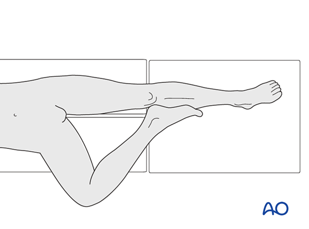
If the patient’s hip is normal, position the patient supine, abduct and externally rotate the leg, and put it in a figure of 4 position. If the hip is stiff position the patient in a lateral decubitus with the involved limb down.
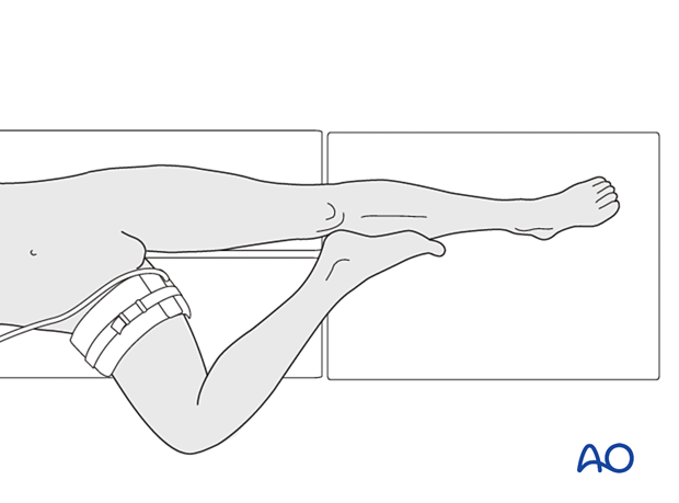
A tourniquet can be considered around the proximal thigh.
2. Skin incision
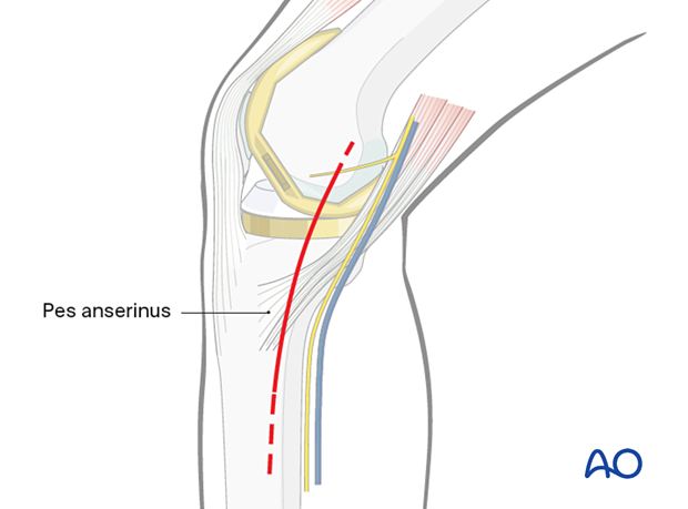
With the knee in slight flexion, make a straight or slightly curved incision running from the medial epicondyle towards the posteromedial edge of the tibia. The incision can be extended as needed both proximally and distally, as indicated by the dashed line.
3. Deep dissection
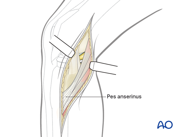
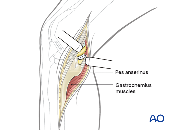
After the opening of the fascia over the semi membranous, identify and expose the remaining pes anserinus.
4. Access
The pes tendons can be reflected posteriorly to expose the posteromedial edge of the tibia, along with the medial aspect of the tibia. Alternatively, the broad insertion of the pes anserinus tendons can be elevated from the anterior without creating instability of the knee.
Caution that the medial collateral ligament is closely adherent to the bone anteriorly and proximally. The MCL should not be elevated from its insertion, as this could lead to knee instability. Plates can be placed directly over the MCL with caution.
5. Wound closure
If needed, insert suction drains and routinely close the soft tissues.


