Medial approach to the distal femur
1. Principles
The medial approach to the distal femur is useful to expose a medial distal femoral fracture. It can also be used when simultaneous medial and lateral plate fixation is performed.
This approach is safe in the presence of a healed previous midline skin incision for a total knee replacement.
2. Skin incision
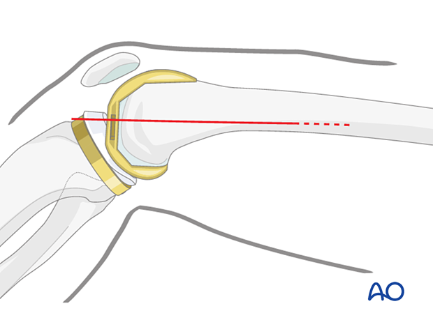
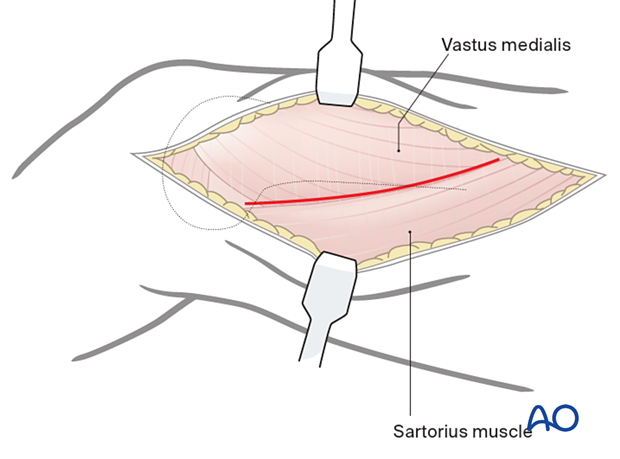
A skin incision is made in the line of the tendon of the adductor magnus. The adductor tubercle is identified, and the line of the adductor tendon is marked proximally. A straight-line incision is made along the posterior border of the adductor magnus tendon. The incision can be extended as far proximally as needed.

3. Deep dissection
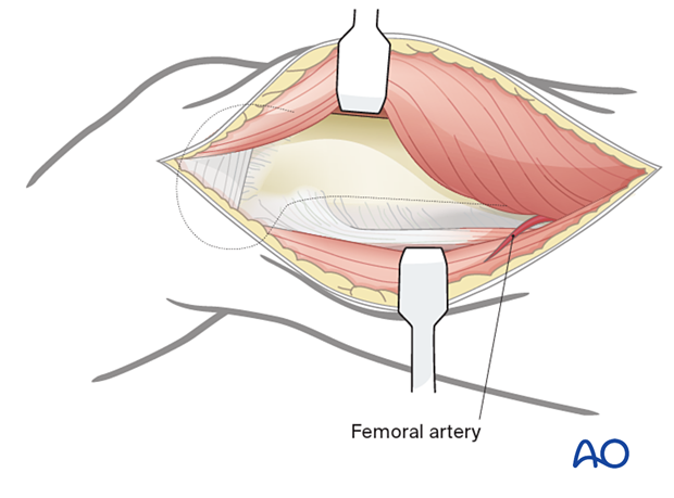
Identify the posterior border of the vastus medialis in the distal portion of the wound. Elevate the medialis anteriorly off the femur moving proximally developing the plane between this muscle and sartorius. As this approach extends proximally, sartorius forms the border of Hunter's canal which contains the femoral vessels. Caution should be taken not to damage the vessels during proximal dissection.

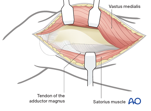
To aid the dissection, flex the knee to allow the anterior border of the sartorius to be retracted posteriorly. This will allow the exposure of the tendon of the adductor magnus. The adductor magnus tendon inserts into the adductor tubercle anteriorly.

4. Exposure
Retract adductor magnus muscle and tendon posteriorly and retract the vastus medialis anteriorly to expose the femur.

5. Wound closure
After careful hemostasis, the wound is closed in layers with absorbable sutures and the chosen skin closure.


