Supine position with manual traction
1. Introduction
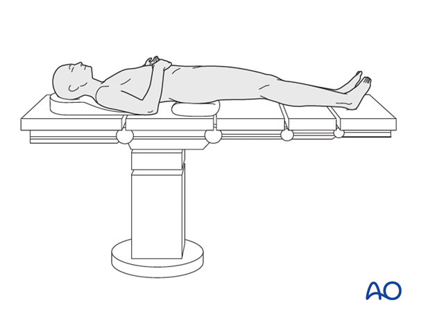
The patient is positioned supine on a conventional table and moved as close as possible to the edge of the table. The pelvis is lifted up by a folded sheet under the ipsilateral buttock.
Note: for periprosthetic fractures, whether performing ORIF or revision arthroplasty, it is preferred to position the patient on a radiolucent table.

2. Preoperative preparation
Operating room personnel (ORP) need to know and confirm:
- Site and side of fracture
- Type of operation planned
- Ensure that operative site has been marked by the surgeon
- Condition of the soft tissues
- Implant to be used
- Patient positioning
- Details of the patient (including a signed consent form and
- appropriate antibiotic and thromboprophylaxis)
- Comorbidities, including allergies
3. Anesthesia
This procedure is performed with the patient under general or regional anesthesia.
4. Positioning
- Reconfigure the table or transfer the patient to a fracture table.
- Reduce the fracture with manual traction and manipulation to ensure reduction is possible before preparing and draping the patient.
- Pad all pressure points carefully (especially in the elderly).
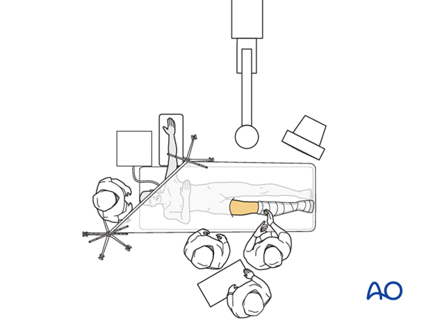
- Position the image intensifier on the opposite side of the injury and the operating surgeon
- Ensure that you can get good-quality AP and lateral x-ray views of the fracture site, full prosthesis, and distal femur before draping.
- In obese patients it may be technically easier to perform the surgery on a lateral position on a radiolucent table.
- Adduct and slightly flex the affected leg anteriorly in front of the unaffected one to ensure the position is reasonable for obtaining X-rays.
- A firm cushion placed in the midline beneath the pelvis may be used to elevate the pelvis from the table edge and facilitate the skin incision.
- The ipsilateral arm should not be positioned on an arm board or abducted, since it could interfere with the surgical procedure. An adducted (pictured) or elevated position is favored.
- The surgeon must be satisfied with the position before the patient is prepared for surgery.

5. Skin disinfecting and draping
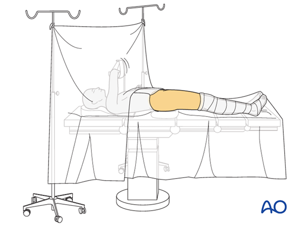
- Maintain light manual traction on the limb during preparation to avoid excessive deformity at the fracture site.
- Disinfect the exposed area from above the iliac crest to the mid-tibia with the appropriate antiseptic.
- Free drape the affected limb(s) with a single-use U-drape. A stockinette covers the lower leg and is fixed with a tape. The leg is draped to be freely moved.
- Drape the image intensifier.

6. Operating room set-up
- The surgeon, assistant, and ORP stand on the side of the injury.
- Place the image intensifier on the opposite side of the injury or surgeon.
- Place the image intensifier display screen in full view of the surgical team and the radiographer.

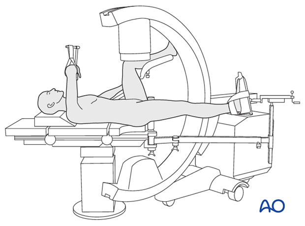
7. Option: use of a radiolucent or fracture specific table
A radiolucent table or a fracture specific table, that can provide traction through the foot and ankle, may be used.
The choice depends on surgeon's experience and preference.


