Lateral approach to the pediatric proximal radius
1. Introduction
The lateral approach can be used to access the lateral condyle, the radial head and the tip of the coronoid process.
2. Skin incision
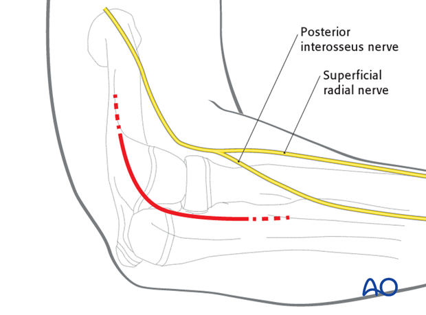
A lateral skin incision is illustrated but a posterior skin incision with a lateral skin flap can also be used.
For a lateral skin incision, place the elbow at 90° and palpate the lateral condyle, which is easier in thin patients.
Make a 5-6 cm gently curved skin incision centered over the lateral condyle, extending proximally or distally if needed.
Note: The posterior interosseous nerve is located within the supinator muscle and must be protected during this approach. This crosses the posterior radius, from anteriorly, three patient finger breadths distal to the radial head.
3. Superficial surgical dissection
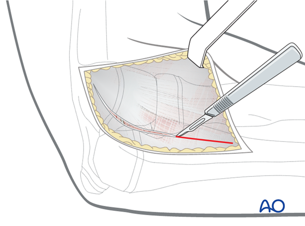
Incise the subcutaneous tissue in line with the incision and raise flaps to expose the fascia over the muscles.
4. Identifying intervals
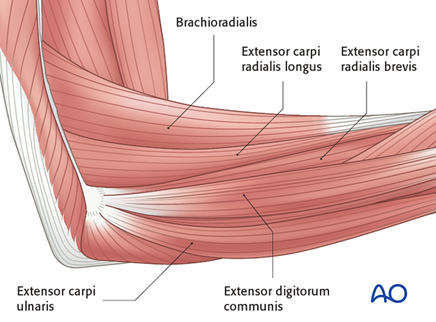
It can be difficult to identify precise intervals proximally because of confluence of fibers in the common extensor origin.
The common extensor origin should be divided in line with its fibers distal to the lateral epicondyle between the epicondyle and the anatomical position of the radial head.
Pronation of the forearm will move the nerve further from the plane of dissection.
5. Avoiding damage to nerves
- Fully pronate the forearm to protect the posterior interosseous nerve by moving it away from the operative field.
- Avoid incising the capsule too far anteriorly as the radial nerve lies over the front of the anterolateral portion of the elbow capsule.
- Avoid dissection distal to the annular ligament or strenuous retraction because the posterior interosseous nerve, lying within the supinator muscle, is at risk.
- Do not place retractors around the radial neck.
6. Deep surgical dissection
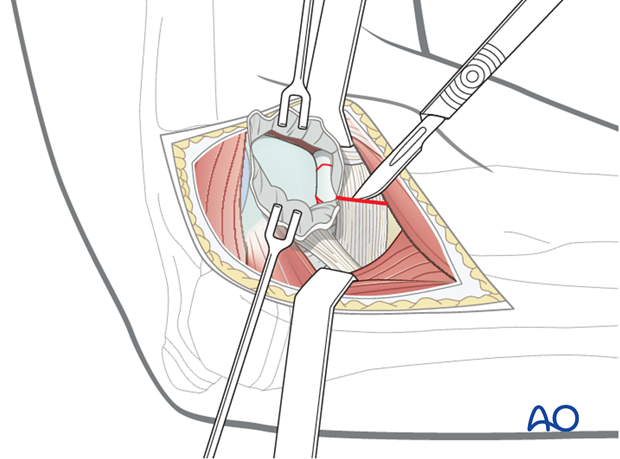
Identify the lateral capsule of the elbow joint and make an arthrotomy proximal to the radial head.
Pearls: To preserve the periosteal vessels first try to reduce the fracture through the closed capsule .
If this is unsuccessful reduce the fracture through the small arthrotomy without dividing the periosteum to the radial head.
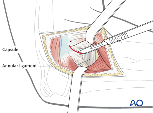
The annular ligament should only be divided if it is obstructing reduction. If required, it may be carefully divided in line with the muscle interval to expose the fracture.
7. Release of proximal capsule and muscle
Release the capsule and muscle from the lateral supracondylar ridge to improve visualization of the capitellum and radial head.
8. Wound closure
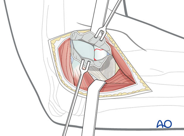
Close the capsule with resorbable sutures (3/0).
Reattach the muscles and fascia with resorbable sutures (2/0 or 3/0).
Close skin and subcutaneous tissue with fine resorbable sutures (this avoids distress to the child when removing nonabsorbable sutures).


