ESIN entry points (pediatric distal radius)
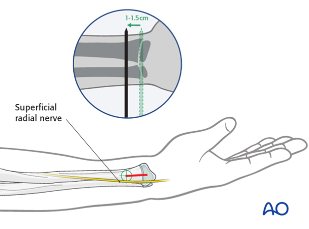
1. Lateral entry point
Using an image intensifier, determine the correct entry point on the lateral side of the radius, 1-1.5 cm proximal to the growth plate.
If an image intensifier is not available, be careful not to injure the growth plate and use an incision, 3-4 cm proximal to the joint.
Start the incision at the level of the lateral entry point and extend it 2 cm distally.

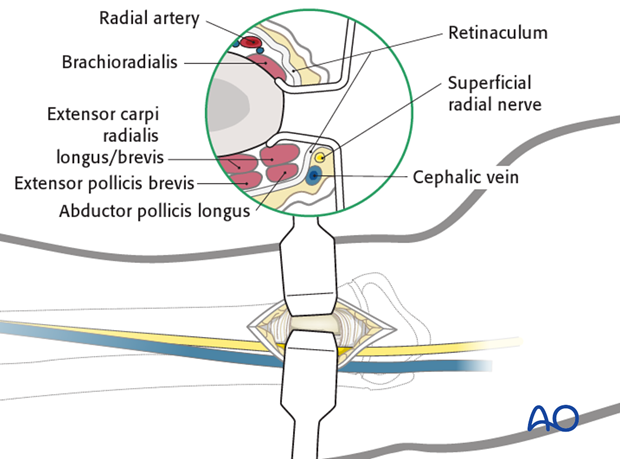
Use small scissors or a surgical clip and small retractors to dissect to bone under direct vision.
Note: Avoid injury to the superficial radial nerve, the cephalic vein and the tendons of the thumb.

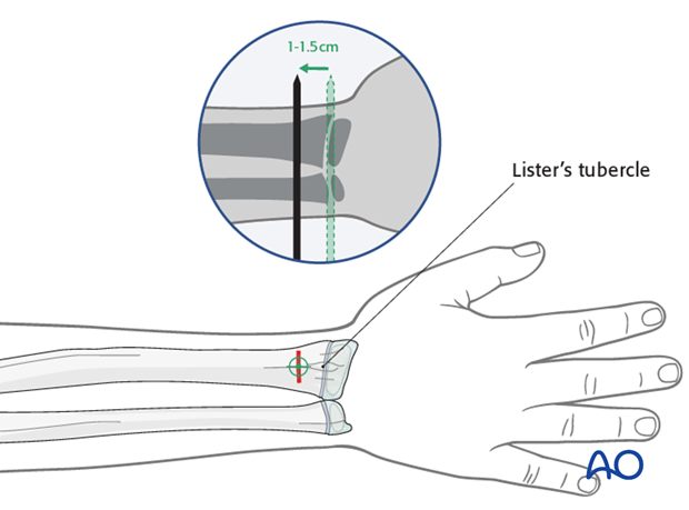
2. Dorsal (Lister’s tubercle) entry point
Use an image intensifier to determine the correct entry point.
This lies in line with Lister’s tubercle, 1-1.5 cm proximal to the growth plate in the metaphysis.
Note: In children the tip of the Lister’s tubercle is part of the epiphysis. Do not open the bone here as it will injure the growth plate. If an image intensifier is not available, use an incision 3-4 cm proximal to the joint or use a lateral entry point.
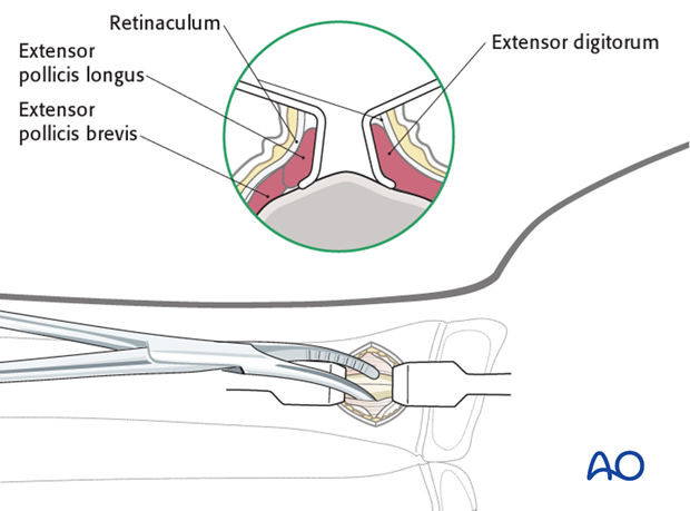
Perform a transverse skin incision along the skin crease. This gives a cosmetic scar and causes less irritation with wrist and hand movement.

Use small scissors or a surgical clip and small retractors to dissect to the bone under direct vision in the following order:
- Skin and subcutaneous incision
- Blunt dissection of the retinaculum
- Blunt separation of the tendons
- Direct visual bone contact

3. Wound closure
Close skin and subcutaneous tissue with fine resorbable sutures (this avoids distress to the child when removing nonabsorbable sutures).


