Classification of children’s fractures
1. Introduction
The AO Pediatric Comprehensive Classification of Long-Bone Fractures (PCCF) was developed by Dr Theddy Slongo and Dr Laurent Audigé and is considered in detail in this section.
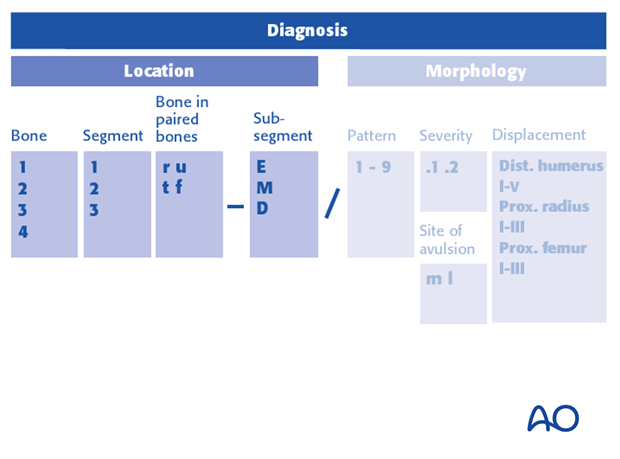
The fracture location comprises:
- The different long bones
- The respective segments
- The subsegments
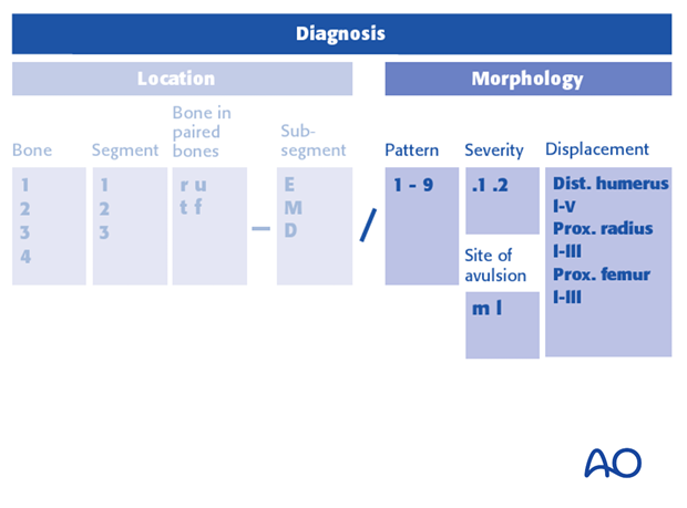
The morphology of the fracture is documented by a specific child code that stands for the:
- Fracture pattern
- Severity code
- Additional code (used in certain types of displaced distal humeral, displaced proximal radial and femoral neck fractures)
2. Location
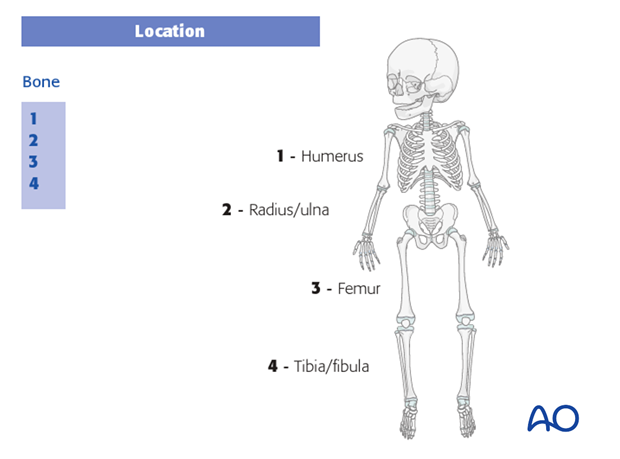
Bone
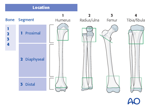
The long bones are numbered:
- Humerus
- Radius/ulna
- Femur
- Tibia/fibula
Segment
Within each of the four long bones, there are three segments:
- Proximal
- Diaphyseal
- Distal
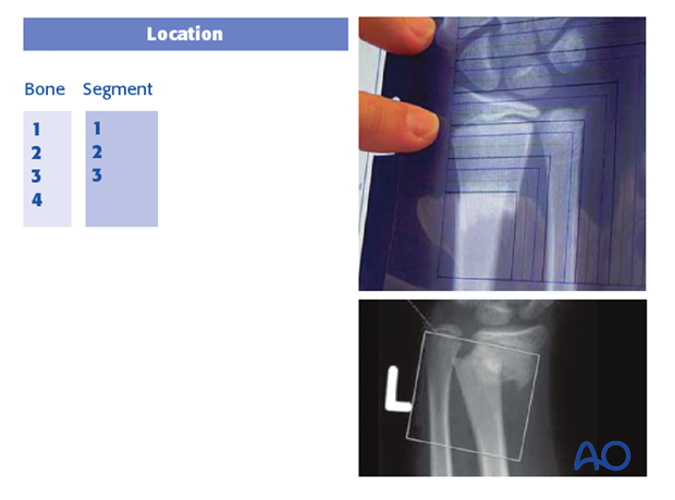
The proximal or distal segment comprises that area of bone covered by a square, the sides of which are equal to the maximal width of the bone at the physeal level.
Paired bones
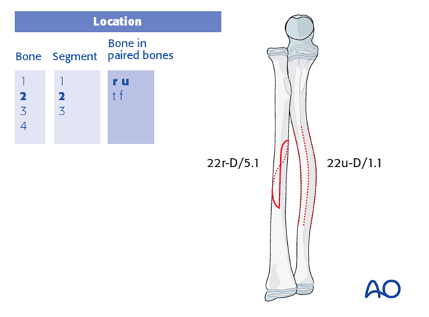
When, in paired bones, (radius/ulna or tibia/fibula) both bones are fractured with the same fracture pattern (see child code), these two fractures should be documented by only one classification code.
In such a case, the severity code will be that of the bone that is more severely fractured.
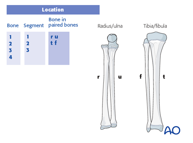
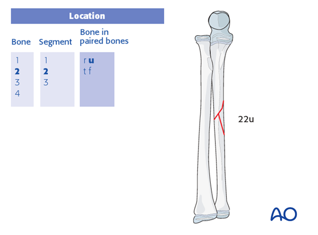
When, in paired bones, only one bone is fractured, a small letter designates this bone (ie, "r", "u", "t", or "f") and should be added to the segment code.
Example: 22u describes an isolated diaphyseal fracture of the ulna.
When, in paired bones, both bones are fractured with different fracture patterns, each fracture must be coded separately.
Example: A complete, spiral fracture of the radius and a bowing fracture of the ulna are coded as 22r-D/5.1 and 22u-D/1.1
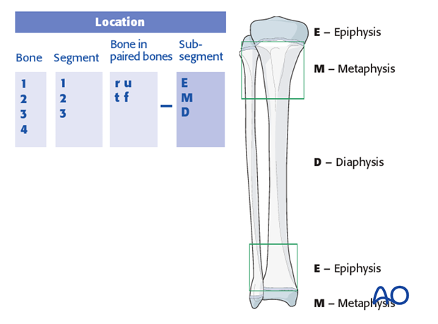
Subsegment
Within the segments 1- and 3- of each diaphyseal bone, the segment is subdivided into E (epiphysis) or M (metaphysis). The diaphyseal segment is coded D.
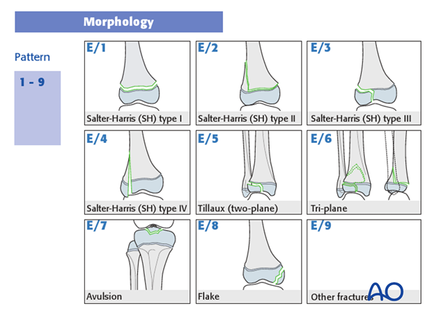
3. Morphology
Pattern
The primary, epiphyseal morphology coding is mapped to the Salter-Harris classification, with some additions as illustrated:
- Salter-Harris type I – E/1
- Salter-Harris type II – E/2
- Salter-Harris type III – E/3
- Salter-Harris type IV – E/4
- Tillaux (two plane) – E/5
- Tri-plane – E/6
- Avulsion – E/7
- Flake – E/8
- Other fractures – E/9
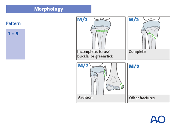
Metaphyseal fractures are coded, as illustrated:
- Incomplete – M/2
- Complete – M/3
- Avulsions – M/7
- Other – M/9
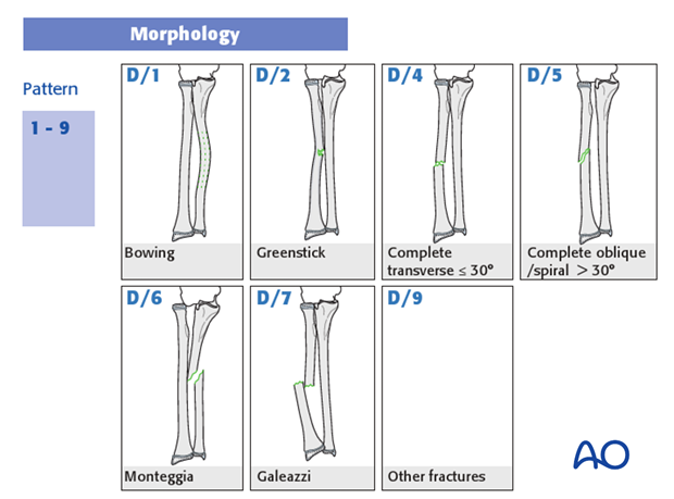
Diaphyseal fracture patterns are coded as illustrated:
- Bowing – D/1
- Greenstick – D/2
- Complete transverse ≤ 30° – D/4
- Complete oblique/spiral > 30°– D/5
- Monteggia – D/6
- Galeazzi – D/7
- Other fractures – D/9
Severity
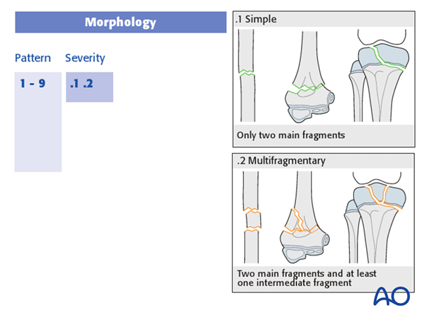
Fracture severity is coded as simple (.1) or multifragmentary (.2).
Site of avulsion
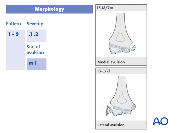
The letters “M” and “L” stand for medial and lateral as modifiers of the M/7 and E/7 avulsion injury codes.
Displacement
Fracture displacement is codified into types, at the distal humerus and the proximal radius.
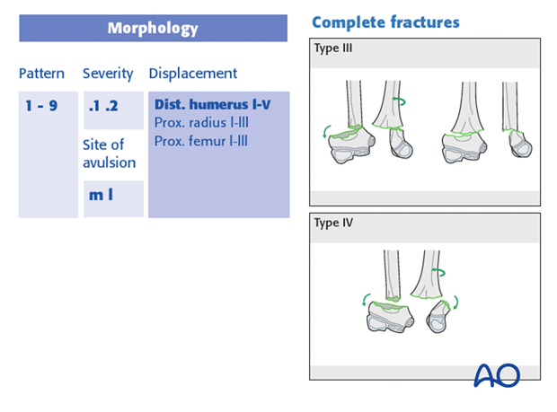
Distal humeral fracture displacements are coded as types I-IV, as illustrated:
- Type I - incomplete fracture where Rogers’ line still intersects the capitellum AND in the AP view there is no more than 2 mm valgus/varus fracture gap
- Type II - incomplete fracture where Rogers’ line does not intersect the capitellum OR in the AP view there is more than 2 mm valgus/varus fracture gap
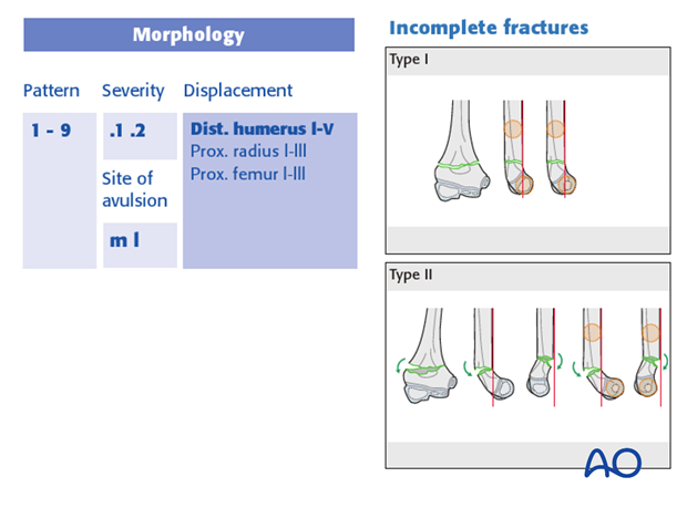
- Type III - complete fracture with no bone continuity (broken cortex), but still some contact between the fracture
- Type IV - complete fracture with no bone continuity and no contact between the fracture surfaces
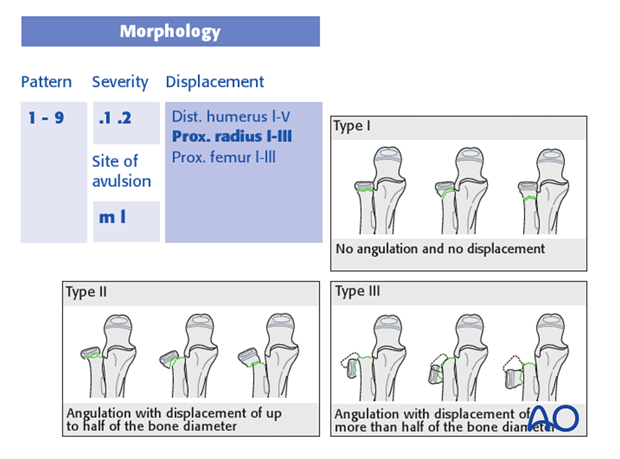
Radial head displacements are codified as Types I-III, as illustrated:
- Type I - no angulation and no displacement
- Type II - angulation with displacement of up to half of the bone diameter
- Type III - angulation with displacement of more than half of the bone diameter
Other modifying codes
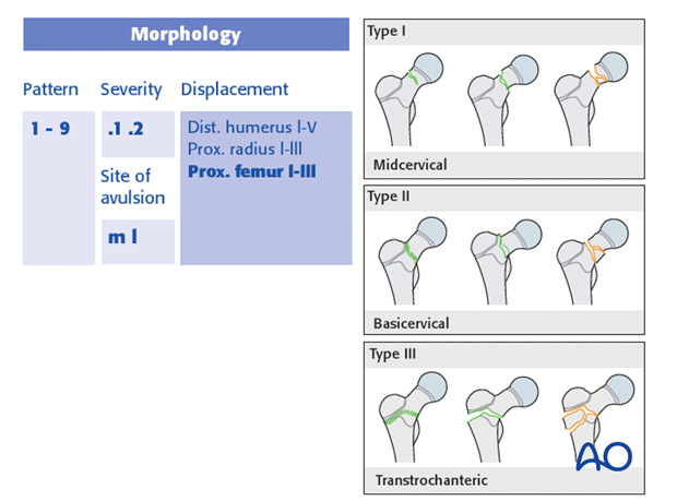
Proximal femoral metaphyseal fractures can be further divided into three types, which are represented by an additional code (I–III) that takes into account the position of the fracture at the proximal metaphysis:
- Midcervical (type I)
- Basicervical (type II)
- Transtrochanteric (type III)
Example: 31-M/2.1 III
4. Further reading
The classification of children’s fractures is covered comprehensively in the illustrated brochure, which can be accessed online as a PDF file.


