Safe zones for pin placement in the pediatric leg
1. Introduction
Inserting percutaneous instrumentation through safe zones reduces the risk of damage to neurovascular structures.
2. Upper leg
Anatomy
The thigh is covered by a circumferential muscular envelope and the diaphysis of the femur by a thick periosteum.
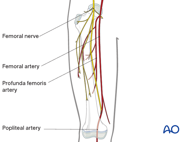
The major neurovascular structures are located medially and posteriorly, and, therefore, the femur can safely be approached over the anterolateral region.
Safe zone in the middle of the shaft
The safest anatomical zones for pin insertion are the anterolateral and direct lateral regions of the femur.
Areas of soft-tissue damage should be avoided, to minimize the risk of subsequent pin-track infection.
See diagrams of cross-sections of the thigh to appreciate the location of muscle groups and neurovascular bundles at each level.
The anatomy in the area between the two solid green lines has a consistent cross-section.
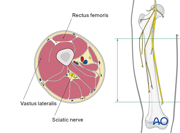
Palpate vastus lateralis and rectus femoris with the patient in the supine position. The direction of the pin should be in the plane between these two muscles, as shown in the cross-section.
Avoid perforation beyond the medial femoral cortex to prevent injury to the neurovascular structures.
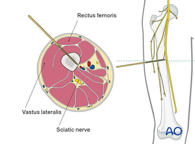
Palpate the vastus lateralis muscle belly and insert the pins in the direction shown in the diagram, aiming to obtain purchase in both cortices.
Avoid inserting pins laterally through the iliotibial tract to reduce the risk of pin loosening and pin-track infection.
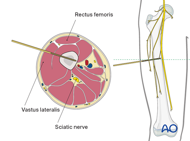
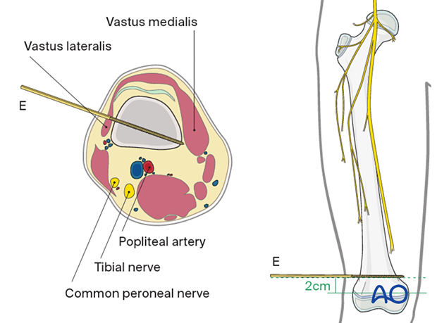
Safe zone in the distal third
Direct lateral approachThe lateral area of the distal part of the femur is easily accessible for pin insertion. The distal part of vastus lateralis is the only structure of the soft-tissue envelope to avoid.
The pin should be positioned at least 2 cm proximal to the growth plate.

3. Lower leg
Neurovascular structures
Common peroneal nerveThe common peroneal nerve runs from the center of the popliteal fossa laterally and curves distally around the fibular head in an anterolateral direction. It separates into a superficial and a deep branch. Injury of this nerve will result in loss of ankle and toe extension, often causing severe functional deficit.
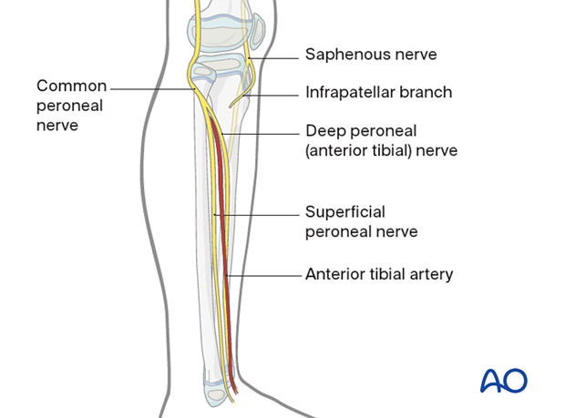
The saphenous nerve runs distally along the anteromedial aspect of the thigh. The infrapatellar nerve branches as it passes the knee joint.
Injury of this nerve will not result in motor deficit but can cause cutaneous sensory loss.
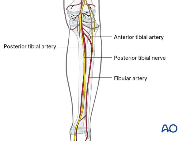
The popliteal artery runs through the center of the popliteal fossa. It separates into the anterior tibial artery, the fibular artery and the posterior tibial artery at the level of the proximal tibial shaft (the trifurcation).
Safe zone in the midshaft of the tibia
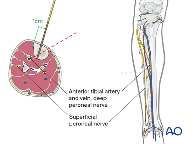
The neurovascular bundle contains the anterior tibial artery and vein, together with the deep peroneal nerve, and runs close to the posterolateral border of the tibia.
These structures are at risk if a pin is inserted in the direction indicated by the red dotted line.
The pins should be inserted approximately 1 cm medial to the tibial crest, on the anteromedial aspect of the tibia, and are angled approximately 20° from the sagittal plane.
