Suture anchors
1. Introduction
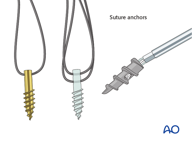
In fractures and soft tissue injuries around the knee, where the bony fragments are too small to fix with screws, the ligamentous and bony fragments can be reduced and fixed in order to obtain a stable knee joint. This can be done with the use of suture anchors.
This procedure may be aided arthroscopically or open.
There are number of different suture anchors on the market which vary in size and design. In this illustration we are using the simple cork screw anchor which is inserted arthroscopically.
2. Patient preparation and approach
Patient preparation
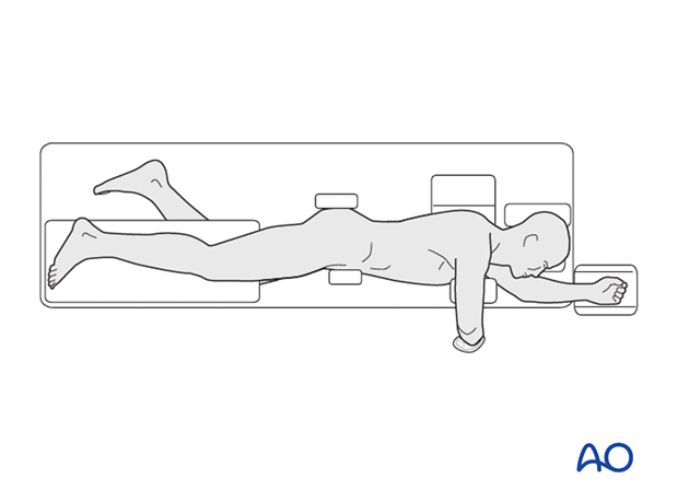
This procedure is normally performed with the patient in a lateral position.
Approach
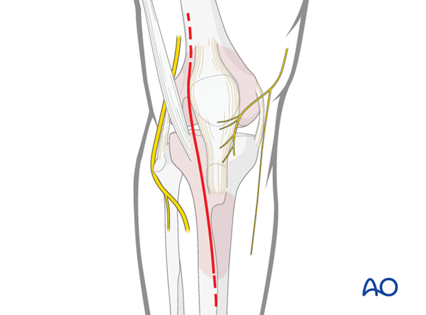
For this procedure an anterolateral approach is used.
3. Anchor insertion
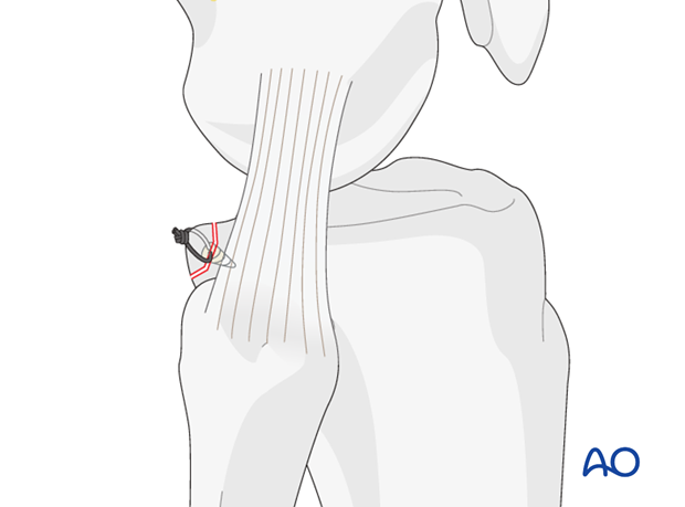
The suture anchor is inserted through the fracture line into bone. The anchors need to be placed sufficiently deep so that the metal part does not protrude above the fracture.
In repairing the soft tissue and small bony fragments, the anchor is placed as close as possible to the articular margin. Once the anchor is securely seated in bone, the handle is removed.
An appropriately angled suture passer is used to shuttle one suture through the labrum.
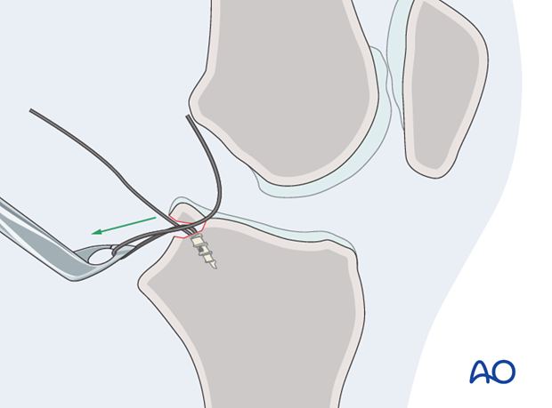
4. Reduction and fixation
The sutures are then tied and as the knot is tightened, the fracture is reduced.
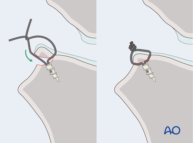
The procedure is repeated for any other fragments not suitable for screw fixation.

At the end of the procedure, use the image intensifier to check the placement of fixation devices and the reduction of the joint.
5. Aftercare
The neurovascular status of the extremity must be carefully monitored. Impaired blood supply or developing neurological loss must be investigated as an emergency and dealt with expediently.
Functional treatment
Unless there are other injuries or complications, mobilization may be performed on post OP day 1. Static quadriceps exercises with passive range of motion of the knee should be encouraged. Early active range of motion of knee and ankle is encouraged.
The goal of early active and passive range of motion is to achieve a full range of motion within the first 4 – 6 weeks. Maximum stability is achieved at the time of surgery. A delay beyond a few days to allow swelling to subside is illogical and harmful.
Weight bearing
A hinged brace is used with non-weight bearing for 6 weeks.
Follow up
Wound healing should be assessed on a short term basis within the first two weeks. Subsequently a 8 week follow-up is usually performed.
Implant removal
Implant removal is optional after retrograde lag screw fixation and suturing.


