Safe zones in the tibia for pin placement
1. General considerations
Make sure to avoid the neurovascular structures around the knee (common peroneal nerve, deep peroneal nerve, and popliteal artery).
Safe zones refers to such placement of thin wires so as to avoid neurovascular structures and intrasynovial placement to minimize the possibility of septic arthritis.
Lower leg
Common peroneal nerve
The common peroneal nerve begins posteriorly in the thigh and runs from the center of the popliteal fossa laterally and anteriorly together and below the tendon of the biceps femoris. It winds anteriorly around the neck of the fibula and then ramifies in the anterior compartment into a superficial sensory and deep motor and sensory branches. Damage to this nerve results in a severe functional deficit.
Saphenous nerve
The saphenous nerve is purely sensory. It runs distally on the anteromedial side of the thigh and passes the knee joint on the medial side of the patella where it gives off the infrapatellar branch. At the ankle it is anterior to the medial malleolus where it runs together with the long saphenous vein. Injury to this nerve will not result in functional deficit but results in a sensory loss and problems with neuroma formation.
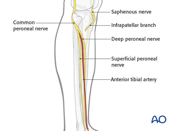
Popliteal artery
The popliteal artery traverses the center of the popliteal fossa. It trifurcates at the level of the proximal tibial shaft into the anterior tibial artery which enters the anterior compartment just above the interroseous membrane, the fibular or peroneal artery, and into the posterior tibial artery.
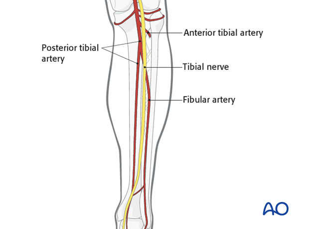
Upper leg
Anatomy
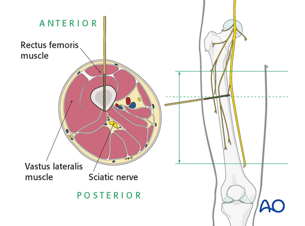
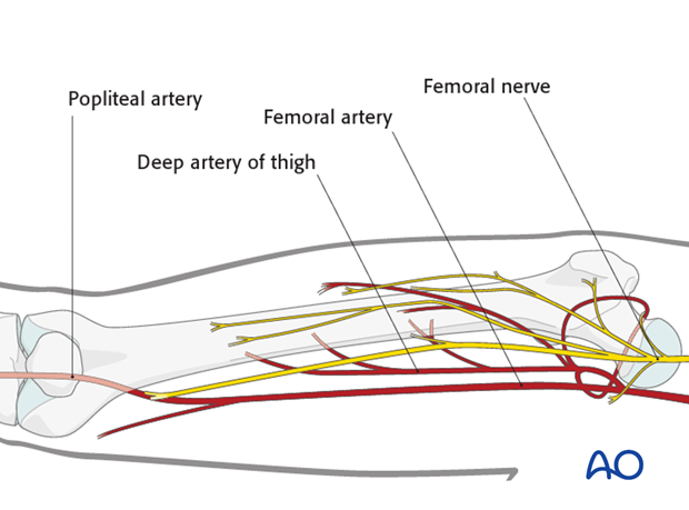
The diaphysis of the femur is surrounded by a thick muscular envelope. The major neurovascular structures are located medially and posteriorly and, therefore, the femur can safely be approached over the anterolateral region.
Overall neurovascular status of the limb
For pin insertion, the condition of the soft-tissue envelope of the femoral shaft has to be considered (areas of crush injuries, or areas of extensive soft-tissue damage should be avoided, in order to minimize the risk of subsequent pin-track infection).
Safe zones
The safest anatomical zones for pin insertion are the anterolateral and direct lateral regions of the femur. See anatomical location of muscle groups and neurovascular bundles in each respective region.
Pin insertion (external fixator)
Locations for pin insertion
The proper locations for pin insertion for external fixators have to be chosen according to the type of external fixator indicated.
Bridging external fixator
For bridging external fixators, the pins are placed anteriorly in the femoral and tibial shafts.
Nonbridging external fixator
For nonbridging external fixators, the pins are placed only in the tibia.
Depth of pin insertion
Pins must be inserted sufficiently deep to engage in the far cortex. In the far cortex the pins must not protrude so as not to injure the neurovascular bundle or the soft tissues.
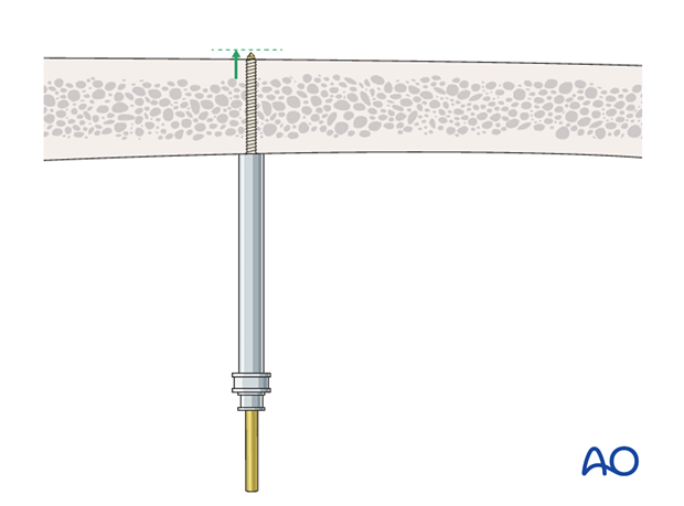
2. Proximal tibia
Proximal tibial head
Neurovascular structures (NVS )
In the depth of the popliteal fossa we find the neurovascular structures in close proximity to the bone. The exact distance of the neurovascular structures to the bone and to the middle of the tibia is variable.
Knee joint capsule
Pin placement should respect the knee joint capsule and therefore be below 2 cm of the tibial plateau. If a more proximal pin fixation is necessary for very high fractures pin placement should be as anterior as possible due to the shorter extend of the knee joint capsule in this area.
See also:
Hutson JJ(2002) Safe Wire and Half Pin Placement in Tibia Fractures,Techniques in Orthopaedics, Lippincott Williams & Wilkins, Inc., Philadelphia, Chapter 2
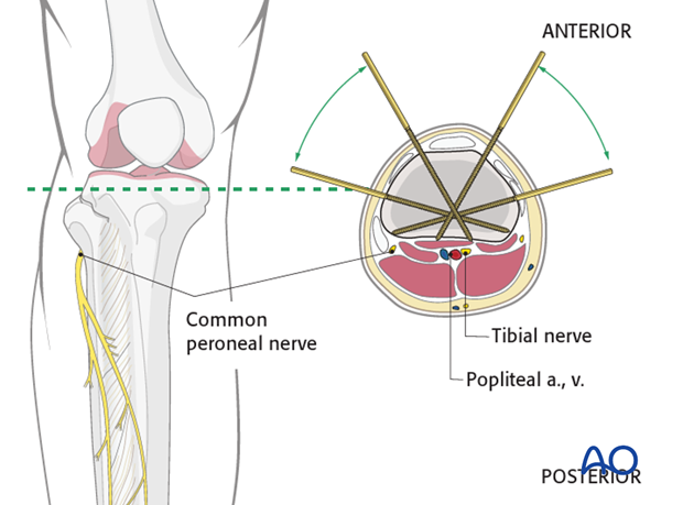
Tibiofibular joint
The common peroneal nerve winds around the fibular neck and then runs anteriorly and distally.
Transfixation
At the level of the fibular head the only safe zones for transfixation of the tibia are the medial and lateral zones.
Unilateral fixation
At the level of the fibular head both sides of the patellar ligament are a safe zone for unilateral frame fixation.
Therefore one can construct a T-frame with good stability with only a minor risk of intra-articular pin placement.
See also:
Bal GK, Kuo RS, Chapman JR, et al (2000) The anterior T-frame external fixator for high-energy proximal tibial fractures,Clin Orthop Relat Res.(380):234-40
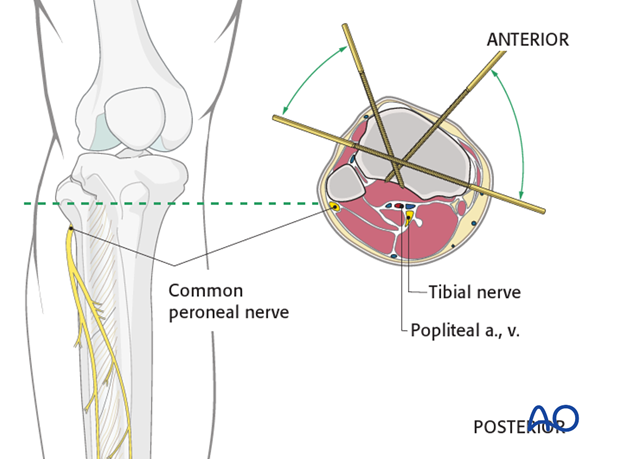
Distal of the tibial tuberosity
To minimize the risk of infection it is best to insert the pins where soft-tissue coverage is minimal.
Therefore distal to the tibial tubercle the safe zones for pin insertion is the tibial crest and the medial face of the tibia.
One must be careful and avoid deep penetration beyond the far cortex.
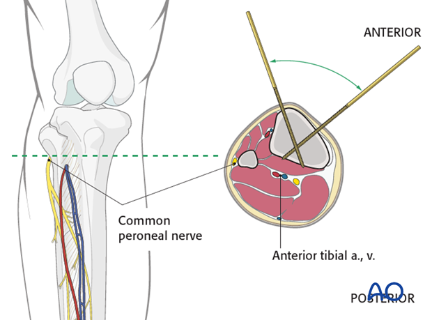
3. Tibial shaft
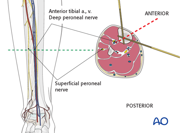
The neurovascular bundle (the anterior tibial artery and vein together with the deep peroneal nerve) run anterior to the interosseous membrane close to the posterolateral border of the tibia.
They are at risk if the pin is inserted in the direction as indicated by the red dotted line approximately half way between the anterior crest and the medial edge of the tibia.
4. Midshaft of the femur
The neurovascular structures in the midshaft of the femur are medial and an anterior placement of the pins will avoid them. The distal pin should avoid the suprapatellar pouch in order to be extraarticular.
