Split fractures of the femoral head
Definition
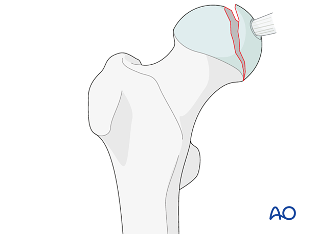
Split fractures of the femoral head are classified by AO/OTA as 31C1.
This fracture type is subdivided:
- 31C1.1 – Avulsion of ligamentum teres
- 31C1.2 – Infrafoveal split fracture
- 31C1.3 – Suprafoveal split fracture (illustrated)
These fractures involve the articular surface. They disturb both the blood supply and the congruity of the articular surface. They are very often associated with hip joint dislocation. The combination with an acetabular (posterior wall) fracture is also frequently seen.

Further characteristics
Associated hip dislocation
In general, fractures of the femoral head are associated with dislocation of the hip. In 90% of cases, the dislocations are posterior and in 10% anterior.
An unreduced dislocation is an emergency because it threatens the blood supply to the femoral head. It may also be accompanied by pressure on a major nerve.
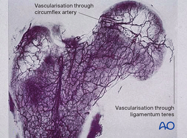
Vascularization through ligamentum teres
The ligamentum teres arises from the transverse acetabular ligament and the posterior inferior portion of the acetabular fossa and attaches to the femoral head at the fovea capitis. Lesions of the ligamentum teres may be caused by dislocation or subluxation of the hip as well as acetabular fractures.
However, the blood supply through the ligamentum teres is minor (10–15% of the femoral head) and mostly to the non-weight-bearing area.

Imaging
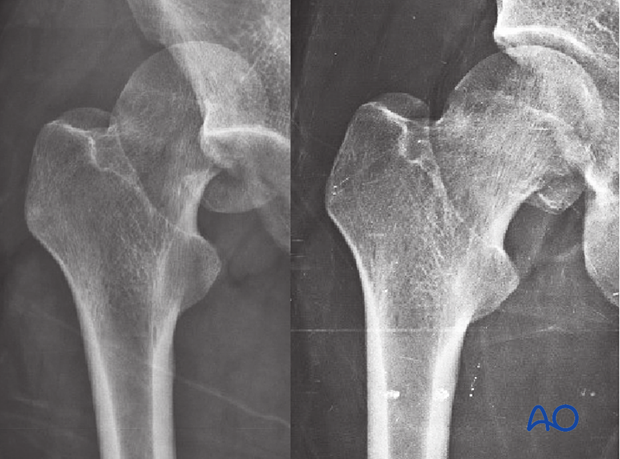
AP and lateral views of the hip and Judet views of the pelvis are needed after closed reduction of the hip. The diagnosis is often missed in case of small fractures, but even larger fractures are not always apparent on a standard pelvic x-ray.
This case shows a femoral head split fracture. On the left x-ray, the dislocation is visible. On the right, the hip is reduced, with the split fragment still displaced.

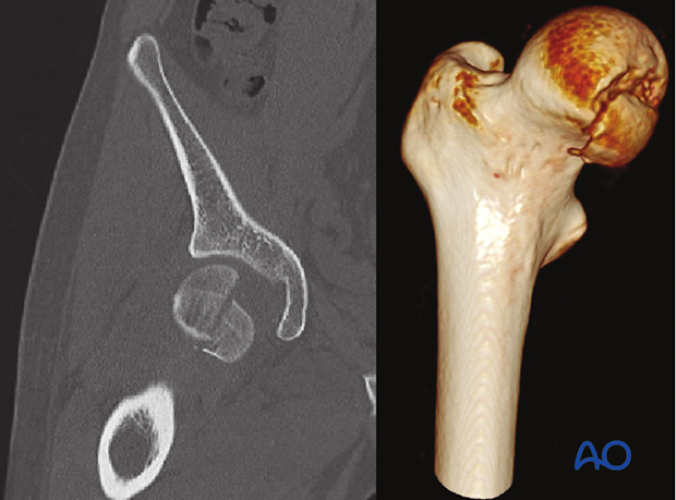
CT is standard imaging as it provides the most accurate information of the fracture.


