Posterolateral (posterior) approach to the hip
1. Preliminary remarks
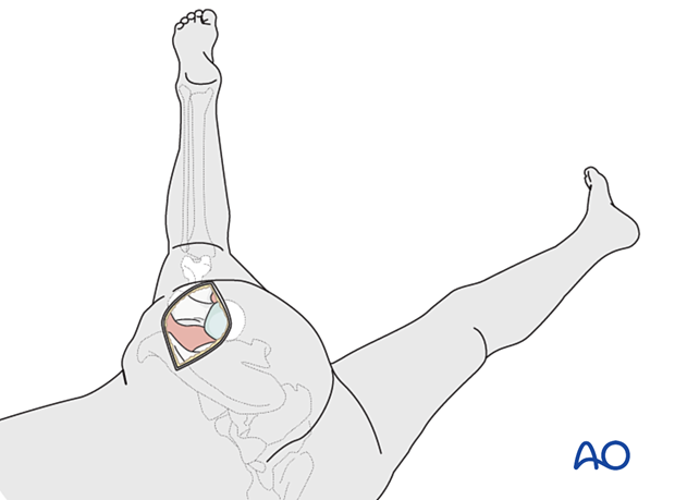
The posterolateral (posterior) approach to the hip is performed with the patient in a lateral decubitus position.
The approach is essentially the same as the Kocher-Langenbeck approach, although done in the lateral position, and the exposure is limited to the hip joint, respecting but not displaying the sciatic nerve. The femoral attachment of the short external rotators and the hip capsule should be repaired to reduce the risk of postoperative dislocation. (Early descriptions of hip arthroplasty through a posterolateral approach suggested excision of the posterior hip capsule.)
For arthroplasty, only the part of the approach proximal to the vastus ridge on the greater trochanter is used (posterior approach).
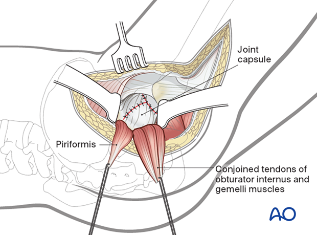
After posterior capsulotomy, the hip is dislocated with internal rotation.
2. Skin incision
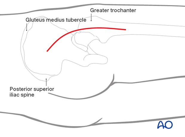
Outline all bony landmarks with a sterile marking pen:
- Posterior superior iliac spine (PSIS), ASIS, and gluteus medius tubercle
- Greater trochanter
- Shaft of femur
Start the skin incision posterior to the lateral side of the greater trochanter and carry it distally about 5 cm along the femoral axis. Proximally, the incision runs slightly curved towards the PSIS to a point approximately 5 cm proximal to the greater trochanter.

3. Dissection of fascia lata
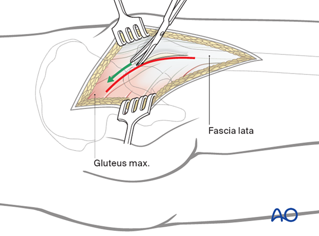
Start sharp dissection of the fascia lata and gluteal muscle at the greater trochanter over the trochanteric bursa. Incise the fascia lata in line with the skin incision.
4. Protection of sciatic nerve
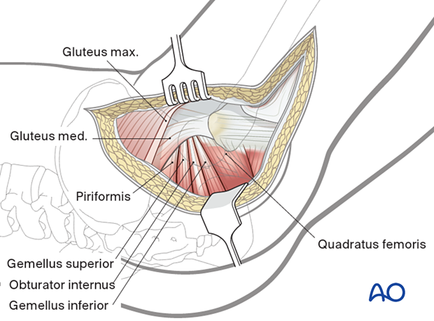
Flex the hip and internally rotate the femur to expose the short external rotator tendinous insertions.
Retraction of the gluteal muscle flap posteriorly shows short external rotators inserting on the femur (at least partially obscured by fat). The sciatic nerve can be palpated posteriorly in the depths of the wound. Its exposure is not necessary for uncomplicated hip arthroplasty, but be aware of the nerve’s location and avoid injuring it with retractors.
5. Exposure of short rotator tendons
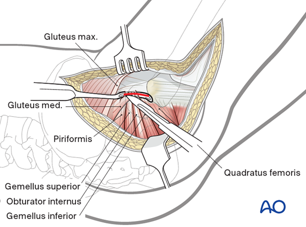
Bluntly dissect the tendinous insertions of the short external rotators and cut them as close as possible to the greater trochanter as any remaining tendinous insertion will be removed when preparing the proximal femur for stem insertion. Place heavy stay sutures for retraction and subsequent repair back to the trochanter. One suture can be placed in the piriformis tendon and another in the conjoined tendons of obturator internus and gemelli.
6. Division and reflection of short rotators
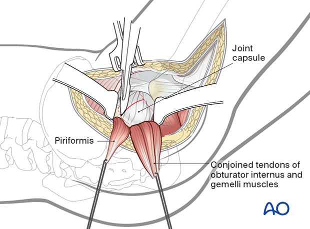
Reflect the short rotator muscles to protect the sciatic nerve and expose the hip capsule.
Enter the joint with a capsulotomy parallel to the femoral neck, with care to identify the acetabular labrum and preserve it when performing hemiarthroplasty.
7. Exposure of hip joint
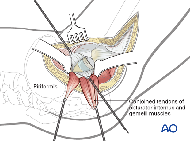
Enlarge the capsulotomy in a T-fashion either proximally or distally as necessary to expose the hip joint.
Create and reflect full-thickness flaps and place heavy nonabsorbable sutures in its free corners to aid in capsular retraction and subsequent repair.
8. Closure
At the end of the procedure, tie the posterior capsular flap sutures. The tendon sutures are passed through drill holes in the posterior greater trochanter and then tied to each other.
Repair quadratus femoris, if divided, separately. A secure repair of the tendons and capsule decreases the risk of hip prosthesis dislocation after a posterior approach.
Close fascia and skin separately.
