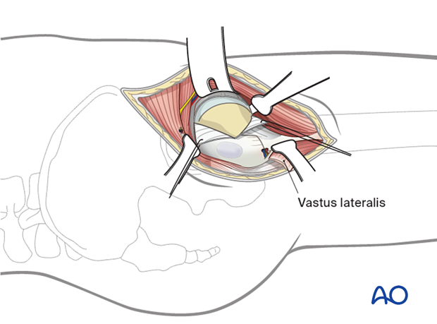
Limited lateral approach for implant insertion in the proximal femur
1. General considerations
The lateral approach is used to insert fixation devices after closed reduction.
The approach provides access to the lateral surface of the proximal femur sufficient for hardware placement.
The incision can be extended proximally or distally as needed.
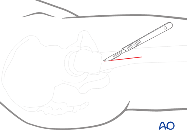
2. Anatomy
The tensor fasciae latae is anterior, between layers of the fascia. Incising the fascia posterior to the tensor avoids injuring this muscle.
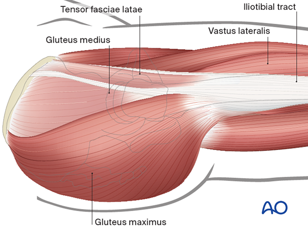
The muscles the surgeon needs to be aware of include:
- Gluteus medius proximal to the greater trochanter
- Vastus lateralis, which covers the femoral shaft distal to the trochanter
No nerves or blood vessels are encountered until the first circumflex artery and vein cross the femur from the posterior 5 cm distal to the trochanter.
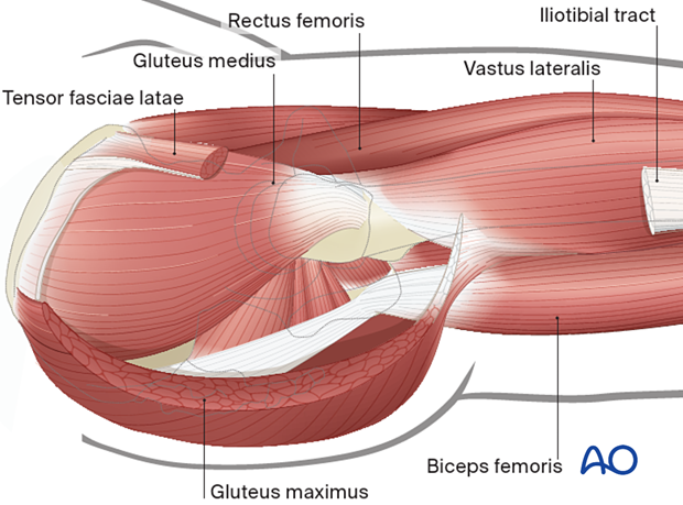
To expose the lateral femoral surface, the vastus lateralis is either reflected anteriorly, if bulky or split longitudinally, if atrophic.
The hip capsule can be palpated anterior to the trochanter.
3. Skin incision
Determining incision placement
The location of the incision depends on the selected fixation device. Screws must be in line with the axis of the femoral neck. A sliding hip screw is also aimed along the center of the femoral neck, but the exposure of the proximal femoral shaft must also accommodate the length of the selected side plate.
Image intensification may be helpful for the proper placement of the incision.
The illustrations show how soft-tissue thickness affects incision placement. Extension of the incision distally is required for the larger thigh.
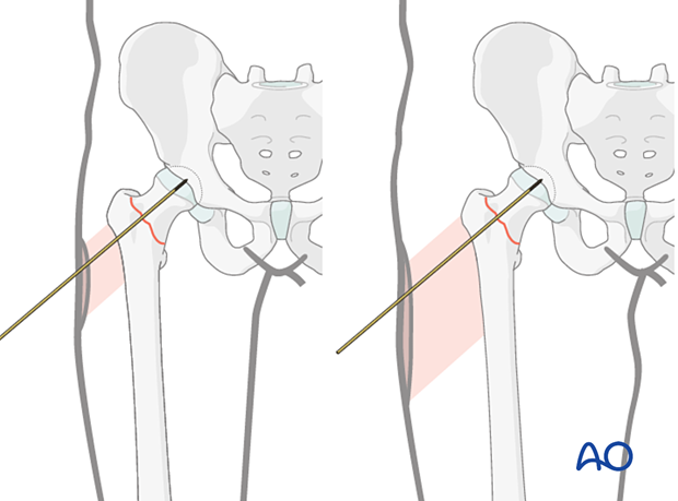
To determine the lateral incision position, overlay the guide wire along the anatomical axis of the shaft; check with image intensification and mark the incision line on the skin.
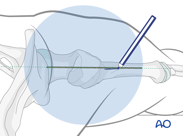
Incision of the skin
For insertion of multiple screws, center the incision over the femoral neck axis line and slightly posterior to the palpable midline of the trochanter.
For a sliding hip screw, the plate angle and length will affect the lateral incision. For example, for a two-hole 135º side plate, the incision usually begins a few centimeters proximal to the palpable greater trochanter and extends 10 cm further distally, over the femoral shaft.
For a 95° angled blade plate, a longer distal incision is required, depending upon the fracture anatomy and length of the plate.
If the vastus lateralis needs to be lifted, extend the incision more distally in patients with bigger muscle mass.

4. Incision of the iliotibial band
Sharply expose the iliotibial band distal to the greater trochanter, and incise it in line with the skin incision.
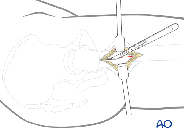
Beneath the iliotibial band, bluntly expose and incise through the fascia of the vastus lateralis. Retracting the mobile muscle mass anteriorly, bluntly divide its fibers to expose the lateral femur.
The first perforating vessels are typically found distal to the location of a short DHS side plate. They should be anticipated if exposure of more than 5 cm below the vastus lateralis ridge (inferior border of greater trochanter) is required.
If divided carelessly, they may cause persistent bleeding.
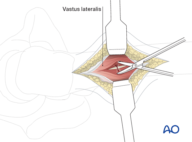
5. Exposure of the femoral shaft
Use one or two small elevators to expose the femoral shaft and place a Hohmann retractor anteriorly. Expose only enough lateral femoral surface for satisfactory hardware placement.
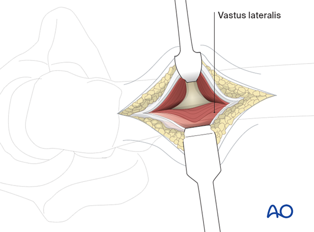
6. Option 1: anterior reflection of vastus lateralis
For muscular patients or increased proximal femoral shaft exposure, the vastus lateralis can be reflected anteriorly by adding a proximal anterior transverse limb to the incision, as illustrated.
Proximal extension of the skin incision aids this variation.
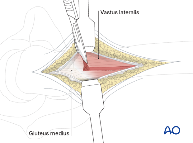
Beginning proximally and posteriorly, elevate the muscle mass from the femur and reflect it anteriorly. The first perforating vessels should be identified and ligated or coagulated.
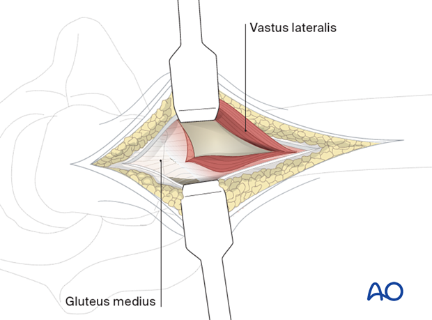
7. Option 2: proximal extension for trochanteric stabilizing plate (TSP)
Proximal extension of the incision through the skin and subcutaneous tissue and fascia lata provides access to the proximal surface of the greater trochanter and insertion of the gluteus medius. This allows sufficient exposure for the surgeon to apply the TSP.
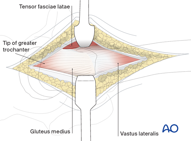
8. Option 3: anterior extension through the hip capsule
For direct access to a femoral neck fracture, the lateral incision can be extended proximally and anteriorly.
Incise skin, subcutaneous tissue, and fascia towards the anterior superior iliac spine. Identify the interval between the gluteus medius and tensor fasciae latae. Retract the gluteus medius posteriorly and the tensor fasciae latae anteriorly; incise the anterior fibers of the hip capsule. Continue this incision onto the anterior lip of the acetabulum. The hip labrum must be preserved. T-shaped capsular incisions allow greater exposure of the femoral neck.
For a complete description, see the anterolateral approach.
