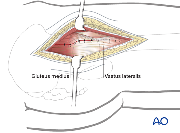
Direct lateral approach to the proximal femur
1. General considerations
The direct lateral approach to the proximal femur releases the anterior third of the gluteus medius and minimus while preserving the posterior femoral attachment of the major part of these muscles. The proximal part of the incision is limited by the superior gluteal nerve and vessels, crossing 3–5 cm proximal to the tip of the greater trochanter. Distally, the anterior fibers of the vastus lateralis are elevated from the anterior femur. The anterior attachment of the hip capsule is next released from the anterior base of the femoral neck, and an anterior longitudinal capsulotomy is opened as necessary with a proximal transverse T-shaped incision.
This approach, usually done with the patient in lateral decubitus position, is excellent for hemiarthroplasty or uncomplicated primary total hip arthroplasty.
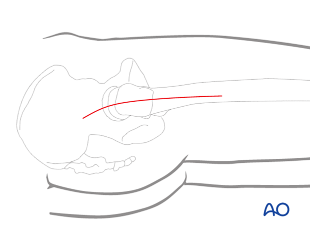
2. Skin incision
Make a longitudinal incision through the skin and subcutaneous tissue, with its proximal end directed slightly posteriorly.
Carry it down to the fascia lata.
3. Superficial surgical dissection
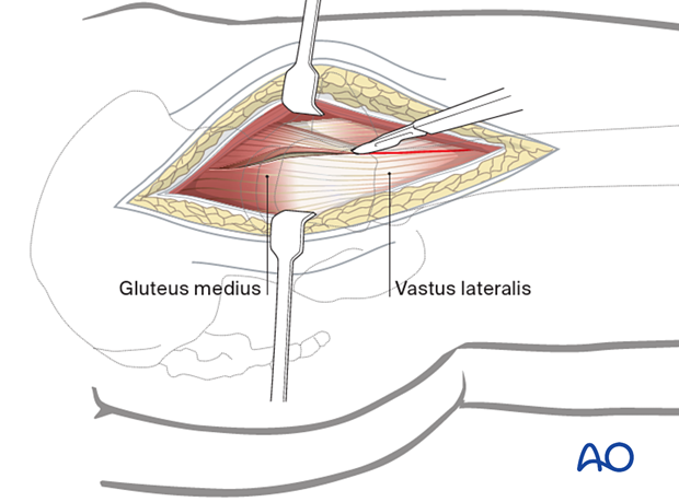
Divide the fascia lata over the greater trochanter, extending it distally over the proximal femoral shaft and proximally splitting the gluteus maximus fibers to reveal the underlying gluteus medius. Remove bursal tissue over the trochanter as needed.
Outline an incision to release the anterior gluteus medius from the greater trochanter. Proximally, this extends into the tendinous insertion of gluteus medius and splitting fibers of vastus lateralis distally. Preserve a substantial portion of gluteus medius insertion posteriorly.
4. Deep dissection
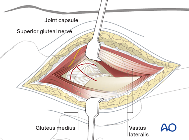
Deepen the incision through the gluteus medius and minimus proximally, retracting the anterior flap to show the hip capsule superiorly and adjacent supraacetabular ilium. Extend the incision distally along the anterolateral femoral shaft and then release the intervening tissue from the anterior intertrochanteric region, sharply releasing the hip capsule from the anterior femur.
Release the capsule sufficiently anteroinferiorly and anterosuperiorly to expose the femoral head and neck and permit free external rotation of the femur. Make a T-shaped capsulotomy to expose the joint, but preserve the acetabular labrum unless a total hip arthroplasty is planned.
5. Hip dislocation
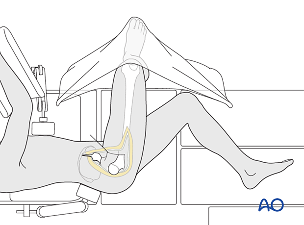
Exposure of the proximal femur is gained by gentle external rotation of the leg. The lower leg is placed into a pocket made from sterile drapes.
Use retractors as necessary to expose the femoral head and neck.
For hip arthroplasty, retraction of the proximal femur distally will allow removing the femoral head fragment from the acetabulum.
6. Wound closure
Close the capsule.
Heavy sutures, typically placed through holes in the bone, are used to reattach the anterior flap to the intertrochanteric region.
Close also the gluteus medius tendon and fascia proximally, and the vastus lateralis fascia distally. Close the fascia lata, subcutaneous tissue, and skin as desired.
