Posterior approach to the SI joint
1. Introduction
The posterior approach to the sacroiliac joint uses and interval between vital anatomic structures.
It provides access to the
- posterior ilium
- sacroiliac joint
- posterior surface of the sacrum
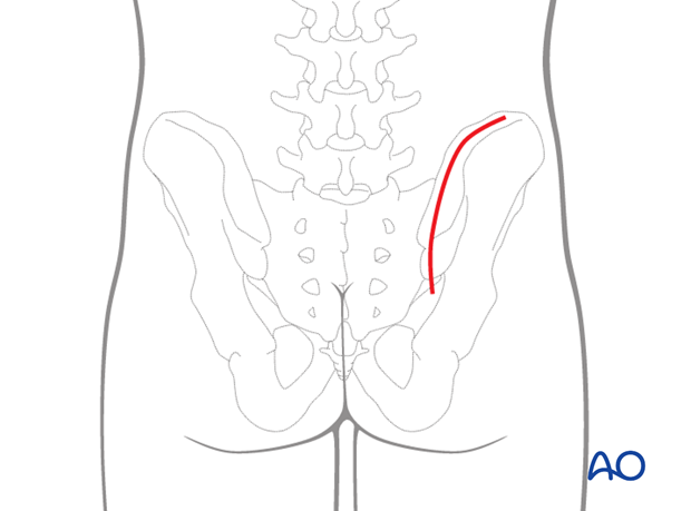
2. Skin incision
Make a curved incision overlying the posterior iliac crest. Begin proximally enough for fracture access and follow the crest distally to the posterior inferior iliac spine.
Note: Posterior approaches through severely contused or crushed tissue or sites of subcutaneous hematoma (Morel- LaVallée lesions) may cause significant wound healing problems.

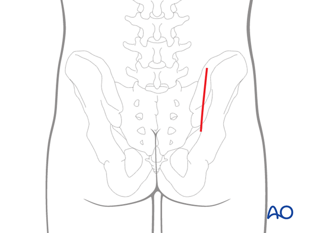
Alternatively a straight vertical incision can be made. The skin incision starts 1-2 fingerbreadths distal and lateral to the posterior superior iliac spine (PSIS) and runs in a vertical straight line proximally (about 10-15 cm in length).
3. Dissection
Divide the subcutaneous tissues in line with the skin incision until the iliac crest is reached.
Reveal the layer of fascia that covers the gluteus maximus muscle and dissect this fascia until the sacral spinous processes are reached.
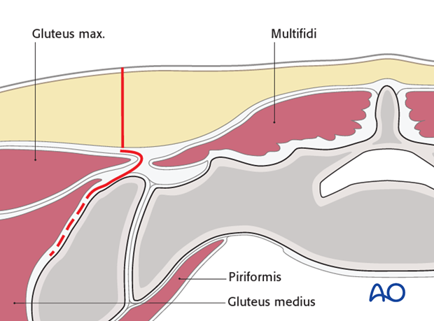
Sharply detach the gluteus muscles from the ilium and elevate them laterally to expose the lateral ilium as needed.
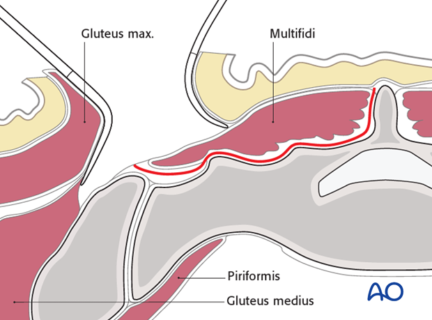
During the subperiosteal exposure of the posterolateral ilium, and elevation of gluteal muscles, it is important to remember the location of the superior gluteal vessels and nerves that are at risk as one dissects anterior and posteriorly.
The muscle cannot be elevated too much anteriorly because their deep surface is tethered by its neurovascular bundle.
The illustration shows the surgeons' finger posteriorly in the sciatic notch where one can palpate the SI joint anteriorly.
Medial to the finger lays the piriformis muscle and then the inferior gluteal neurovascular bundle.
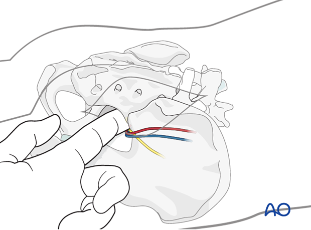
The inferior gluteal artery and vein, along with the inferior gluteal nerve, exit the greater sciatic foramen at the caudal edge of piriformis, and then run upwards to enter the deep surface of the gluteus maximus, to provide its blood circulation and enervation.
Protect them from sharp injury, and excessive tension, during this exposure. Note that they wrap around the piriformis, and prevent its being mobilized distally.
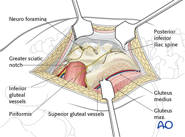
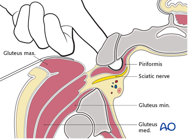
SI joint alignment can be assessed by palpation through the greater sciatic notch, with a finger dorsal to the piriformis and anterior (ventral) to the SI joint. The joint edges should be level, and without a gap.
4. Closure
Reattach the gluteus muscles to the medial fascia flap, over the posterior ilium.
Close the subcutaneous tissues and skin in layers.


