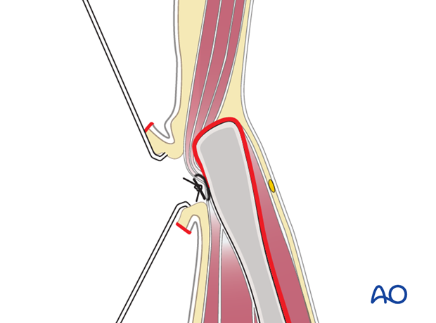
Anterior approach to the iliac wing and SI joint
1. Indications
The anterior approach to the iliac wing and the sacroiliac joint is useful for surgical exposure to manage:
- Sacroiliac joint dislocations
- Fractures of the ilium, even extending into the sacroiliac joint
This approach enables direct visualization of the anterior and superior portions of the sacroiliac joint.
2. Access
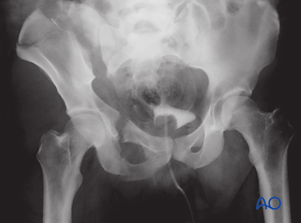
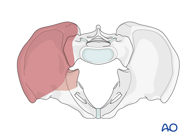
This anterior approach provides access to the, iliac crest, the entire internal iliac fossa, and the most lateral sacral ala. This is limited by the L5 nerve root on the anterior aspect of the ala.
The exposure incorporates full visualization of the anterior aspect of the sacroiliac joint, if needed.
The approach also provides digital and limited visual access to the quadrilateral surface and greater sciatic notch.
3. Potential risk
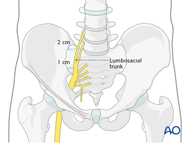
The L5 and L4 nerve root (lumbosacral trunk) travels across the sacral ala and is 10-15 mm medial to the sacroiliac joint.
Because of the proximity of this lumbosacral trunk, the surgeon must use care when working in this area.
Note
Sacral fractures cannot be exposed through this approach because of the overlying nerves and vessels.
4. Skin incision
An incision is made along the iliac crest. Be aware of the lateral femoral cutaneous nerve in the region of the ASIS.
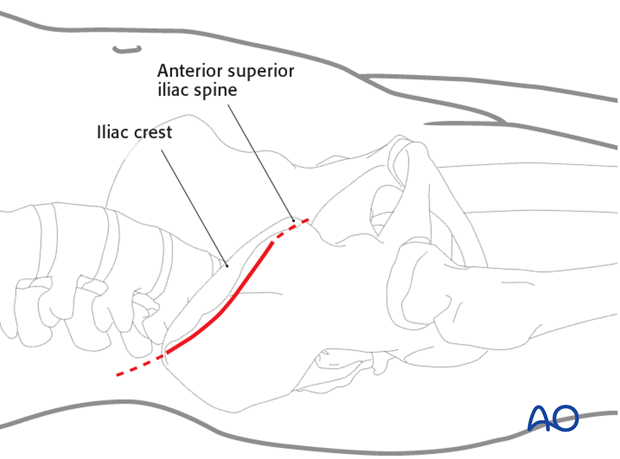
The length of the incision is determined by the amount of area that needs to be accessed in the iliac fossa.
The incision can be extended intraoperatively depending on the necessary exposure.
For fractures involving the posterior aspect of the ilium, or the sacroiliac joint, the exposure needs to be extended posteriorly almost to the table.
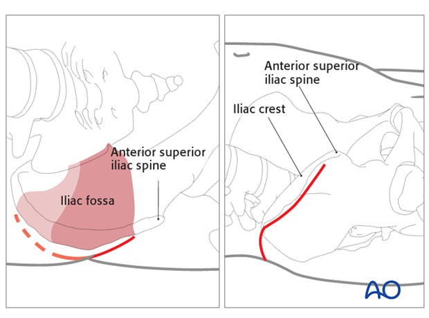
5. Superficial surgical dissection
Divide the subcutaneous tissues in line with the skin incision in order to expose the fascia overlying the external oblique muscle.
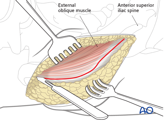
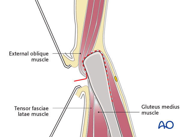
Identify the border between the gluteus muscles and external oblique muscles. Incise the muscular interval with electrocautery.
The external oblique muscle is subperiosteally elevated from the iliac crest.
With a small elevator, the iliac muscles are elevated using the same subperiosteal layer.
6. Deep dissection
When elevating the iliacus muscle, bleeding from nutrient vessels can occur and should be stopped with bone wax.
Continue with careful blunt dissection to the interior part of the SI joint medially to the pelvic ring.
Proceed anteromedially at the pelvic rim as far as to where the iliopectineal eminence begins.
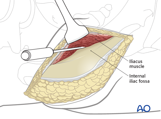
Continue the dissection with an instrument such as a Cobb elevator.
The sacroiliac joint capsule should be identified. A portion of the capsule may be disrupted in the case of sacroiliac joint injury.
Place a Hohmann retractor into the superior portion of the sacroiliac joint.
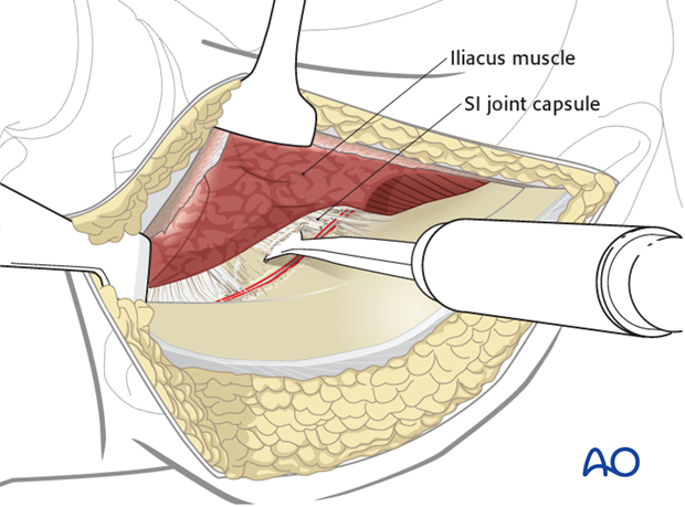
7. Expose the SI joint
The dissection is carried further medially onto the sacral ala. Move the Hohmann retractor medially carefully onto the sacral ala.
Note the L5 nerve root traversing this region.
Place a second Hohmann more anteriorly on the sacral ala.
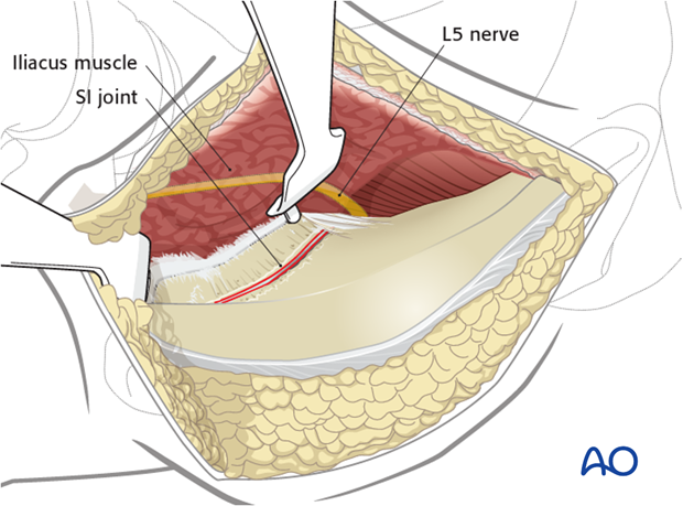
Place an additional Hohmann anterior to the sacroiliac joint for additional exposure. Take care when placing this anterior retractor as it may injure the superior gluteal artery and nerve as they exit the greater sciatic notch.
The sacroiliac joint is now exposed.
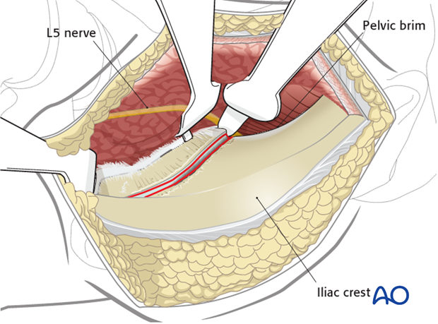
8. Wound closure
Repair the aponeurosis of the external oblique muscle to the periosteum that was left intact lateral to the iliac crest.
Close the subcutaneous tissues and skin in layers.
