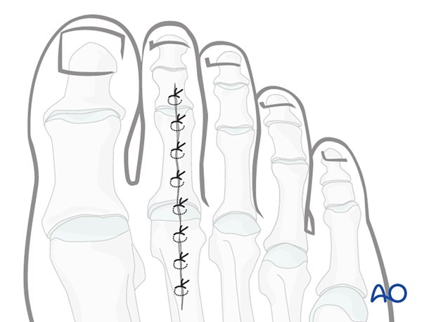
Dorsal approach to the proximal phalanx
1. Indications
This approach is indicated for fractures of the proximal phalanx that requires ORIF.
It can also be used for flexor tendon transfer to the first phalanx, PIP arthrodesis, MTP release, and/or extensor digitorum longus or brevis release or lengthening.
2. Anatomy
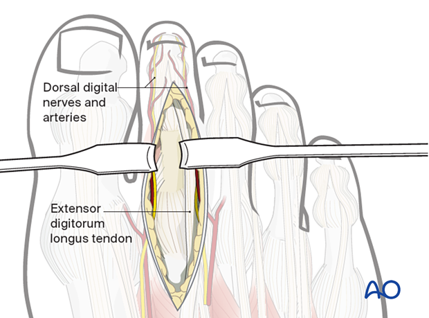
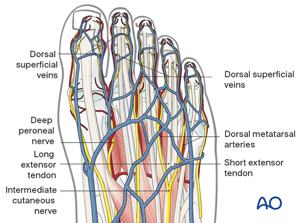
The veins are superficial and should be preserved, especially those which run longitudinally in the long axis of the toe.
As arteries and the nerves are mainly situated on the medial and lateral aspects of the toe, the best approach is in the sagittal plane, in line with the dorsal axis of the toe.
3. Skin incision
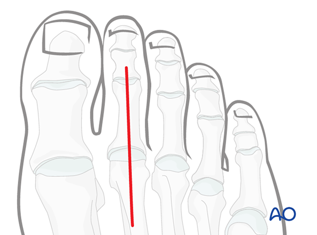
Make a longitudinal incision over the center of the toe from the MTP joint up to the PIP joint.
4. Division of the extensor tendon
Avoid cutting the longitudinally running veins.
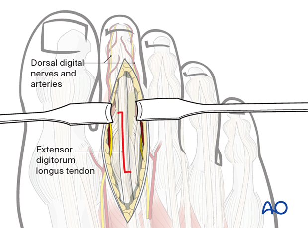
Expose the extensor tendon and retract it or…

….incise it in a Z-shape fashion. Make a short transverse cut proximally and distally.
One of the cuts should be made from the midline laterally and the other from the midline medially.
Then join the two cuts with a longitudinal midline cut.
5. Capsulotomy
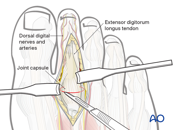
Expose the capsule. Dissect out the sides of the joint capsule using scissors, and insert small right-angle retractors to protect the neurovascular structures on each side of the capsule.
Make a cut ¾ of the perimeter of the capsule.
Do not cut the volar plate.
6. Wound closure
The capsule may be left open if a repair is not possible.
If divided, repair the tendon using a 3.0 suture.
Close the wound with a running 4–0 self-absorbing suture.
