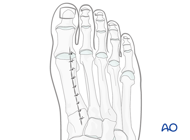
Dorsal approach to the 1st metatarsal
1. Indications
The dorsal approach to the first metatarsal is used to access the entire 1st metatarsal.
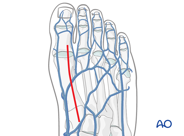
2. Anatomy
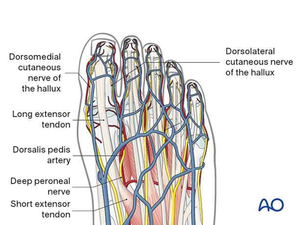
Important, relevant anatomical structures are:
- The extensor hallucis brevis and longus
- The deep peroneal nerve and its branches
- The dorsalis pedis artery
- The dorsomedial cutaneous nerve of the hallux
- The digital branch to the second toe
3. Skin incision
The skin incision is made in line with the first ray, starting over the medial cuneiform and extending distally.
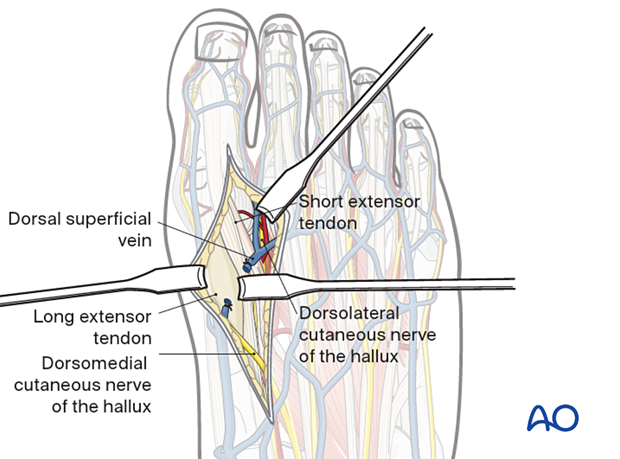
If the metatarsal phalangeal joint is to be exposed, the incision is performed medial to the EHL tendon, between the branches of the deep peroneal nerve, the superficial peroneal nerve, and the saphenous nerves. If needed, it can be extended proximally up to and across the first TMT joint.

4. Deep dissection
Protect any longitudinal veins traversing the field.
Proximally, take care to dissect medial to and not damage the dorsalis pedis vessels and the cutaneous branches of the deep peroneal nerve.
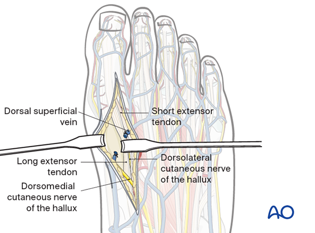
Retract the EHL tendon laterally and expose the first metatarsal.
Visualization of the articular surface
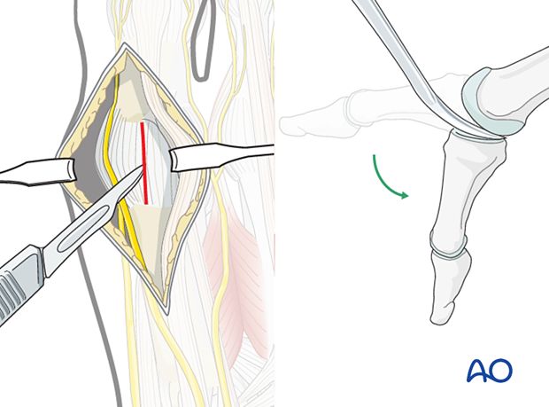
Incise the joint capsule to expose the first metatarsal head and base of the proximal phalanx.
If additional exposure is required, release the medial and lateral collateral ligaments sharply from the first metatarsal head. Free the plantar sesamoids with a smooth rounded elevator. Plantar flex the great toe to visualize the articular surfaces of the joint. A mini distractor applied dorsomedially may be helpful for visualization.
5. Closure
Close the joint capsule using resorbable sutures.
Repair the tendon sheath over the extensor hallucis tendon if possible.
Perform a subcuticular closure with resorbable sutures.
The skin should be closed with appropriate sutures. Nylon sutures are typically used.
