Supine patient position
1. Introduction
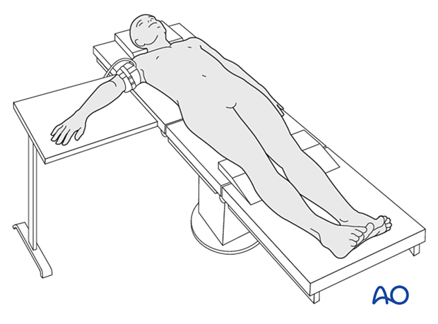
The patient is usually in a supine position with the arm on a radiolucent hand table.
2. Preoperative preparation
For surgery, the operating surgeon needs to know:
- Details of the patient (including a signed consent form and appropriate antibiotic and thromboprophylaxis)
- Patient positioning
- Comorbidities, including allergies
- Site and side of the fracture
- Ensure that the operative site is marked
- Type of operation planned (percutaneous or open approach)
- Implants to be used
- Condition of the soft tissues
3. Anesthesia
- General anesthesia
- Regional nerve block
- Combination of nerve block and light general anesthesia
Prophylactic antibiotics are optional.
Alternatively, the patient stays awake with local anesthesia (WALANT) without a tourniquet. This allows active movement of the hand and fingers during the operation, especially to check for alignment.
- Lalonde DH, Wong A. Dosage of local anesthesia in wide awake hand surgery. J Hand Surg Am. 2013 Oct;38(10):2025–2028.
- Joukhadar N, Lalonde DH. How to Minimize the Pain of Local Anesthetic Injection for Wide Awake Surgery. Plast Reconstr Surg Glob Open. 2021 Aug4;9(8):e3730.
- Lalonde DH. Wide awake hand surgery. Stuttgart, New York: Georg Thieme; 2011.
4. Patient positioning
Position the patient supine and place the forearm on the radiolucent hand table.
As the hand will tend to be semisupinated internally, rotate the shoulder to allow the hand to be placed in an appropriate pronated position for a dorsal approach.
Supinate the forearm for a palmar approach.
In unusual circumstances, a combined dorsal and palmar approach may be required.
A pneumatic tourniquet is recommended.

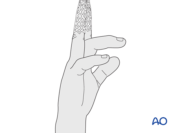
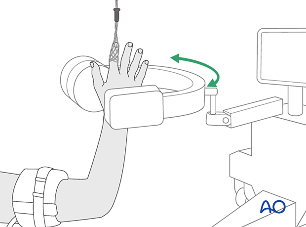
The affected finger may be suspended in a finger trap to apply traction, assist in reducing fragments and/or dislocations, and permit arthroscopic approaches.
C-arm positioning
The image intensifier should be positioned to not interfere with the operating surgeon’s access to the surgical field.
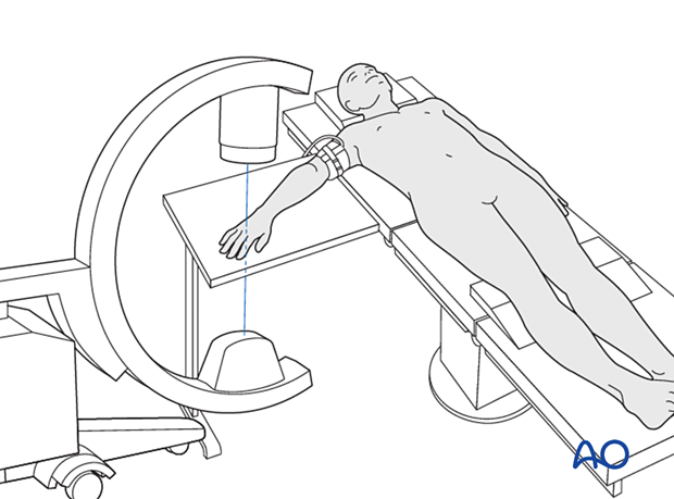
If a finger trap is used, a small image intensifier may be helpful.
Skin disinfecting and draping
Disinfect the entire hand, wrist, and arm with the appropriate antiseptic right up to the limits of the tourniquet cuff. This allows full exsanguination.
Preparation of the entire upper limb allows repositioning during surgery.
A protective specific tourniquet exclusion drape can be used to avoid tracking of antiseptic solution under the tourniquet while in use: if alcohol-based antiseptic is used, skin damage can occur from prolonged contact with soaked material during surgery.
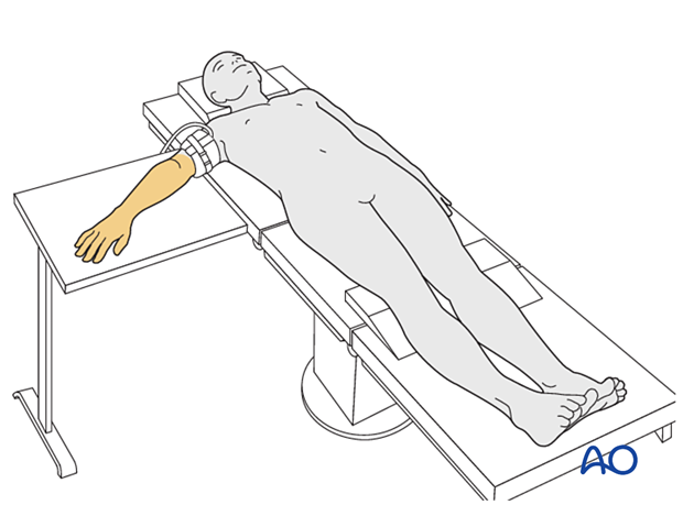
A single-use occlusive hand drape with an expandable arm opening is recommended to isolate the upper limb.
Drape the image intensifier.
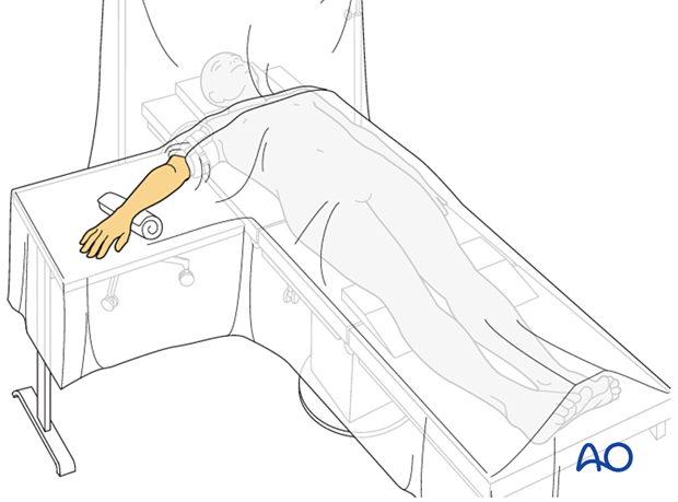
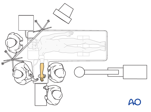
Operating room set-up
The surgeon sits beside the patient’s head to gain a good view and access to the dorsum of the hand. If a volar approach needs to be performed, the surgeon sits on the opposite side of the arm table.
The assistant sits opposite the surgeon. The ORP sits at the end of the hand table.
Place the image intensifier screen in full view of the surgical team and the radiographer.
