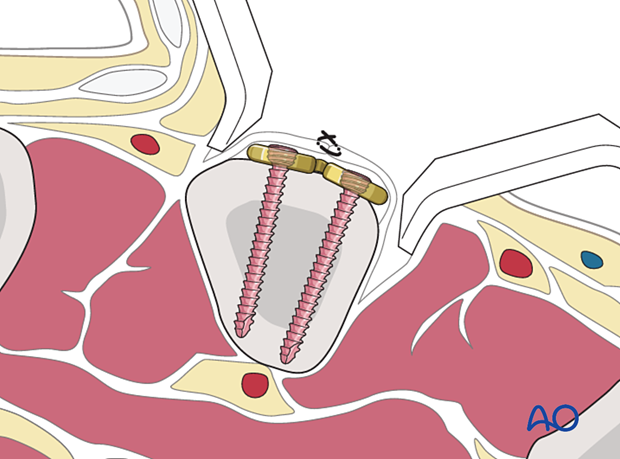
Radial approach to the 2nd metacarpal
1. Indications
This approach exposes the distal 2/3 of the 2nd metacarpal.
It is indicated for open treatment of the following fracture types of the diaphysis and metaphysis:
- Oblique
- Spiral
- Transverse
- Multifragmentary
It can also be used for corrective osteotomies of malunited fractures.
This approach avoids conflict with the extensor mechanism of the metacarpophalangeal (MCP) joint.
2. Surgical anatomy
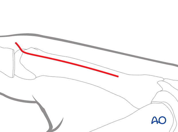
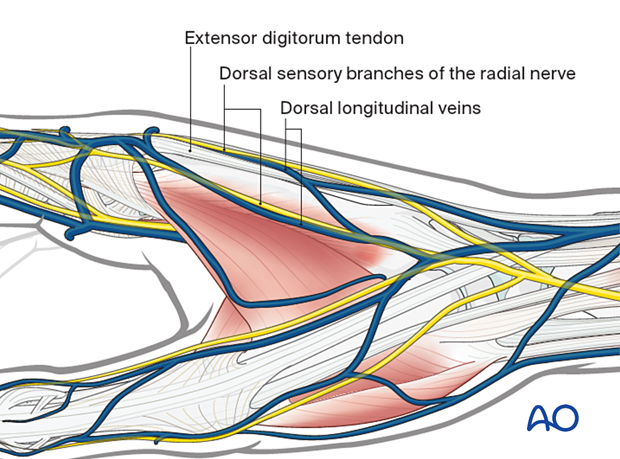
The radial aspect of the 2nd metacarpal can be approached easily, as the two extensor tendons of the index finger run slightly obliquely toward the center of the wrist joint.
3. Skin incision
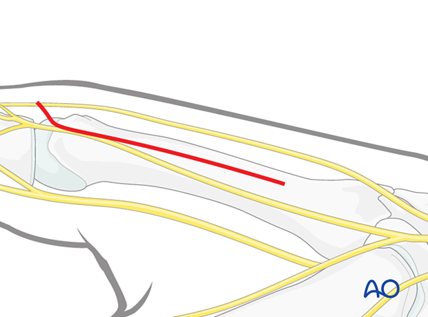
Start the incision dorsal to the center of rotation of the MCP joint. Continue with a straight longitudinal skin incision radial to the diaphysis.
4. Retraction of extensor tendons
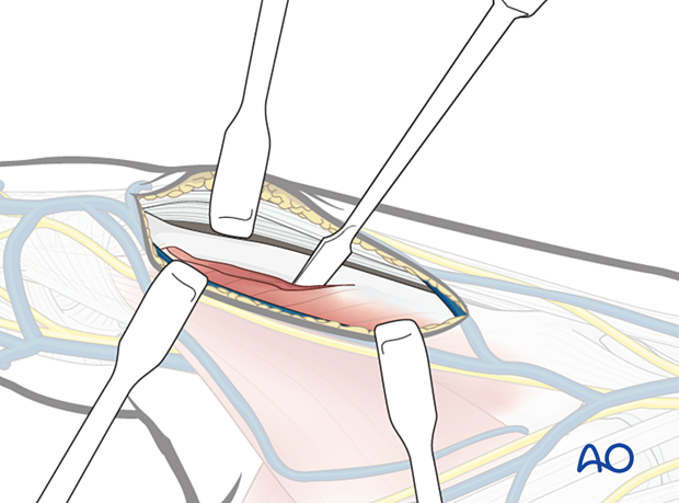
The extensor tendons can be retracted to the dorsoulnar side, together with the surrounding loose connective tissue.
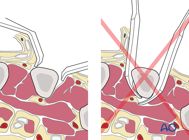
If necessary, partially detach the dorsal interosseous muscles subperiosteally from the bone.
Take care to protect the radial cutaneous sensory nerve during dissection.
If exposing the metacarpal neck, protect the sagittal bands.
Pitfall: Avoid complete muscle detachment
5. Wound closure
Cover the implant with the periosteum as far as possible; this helps minimize contact between the extensor tendons and the implant.
