Posteromedial approach to the malleoli
1. Indications
This approach is indicated in cases of posterior comminution and/or a posterior extension of a medial malleolar fracture.
2. Incision
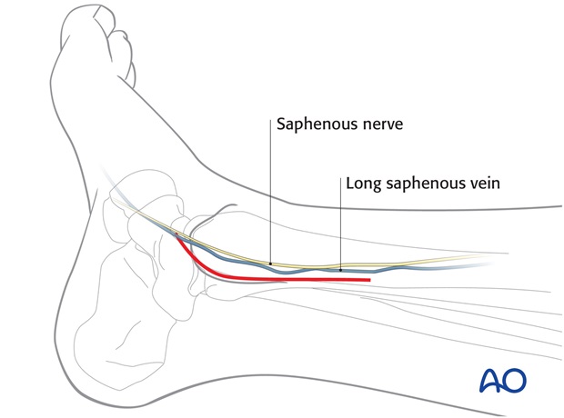
Start the incision 1 cm distal and 1 cm anterior to the middle of the tip of the medial malleolus. Curve the incision dorsally over the tip of the medial malleolus and in the direction of the posterior crest of the distal tibia.
Note
Be careful not to damage the saphenous vein and nerve, especially distally.

3. Superficial surgical dissection
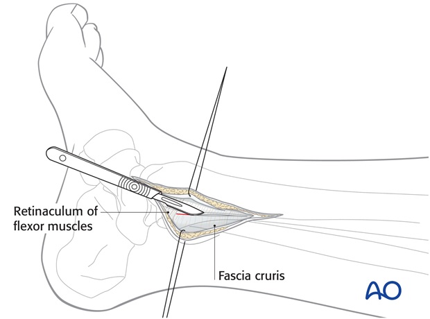
Deepen the approach through the subcutaneous fat and the fascia, in a direct line with the posteromedial crest of the tibia.
Deepen the dissection of the fracture site without stripping off the periosteum.

4. Deep surgical dissection
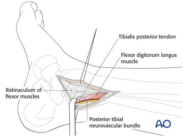
Follow the fracture line to the posterior edge of the distal tibia. Open the crural fascia at the edge of the posterior tibia, proximally as far as necessary, and distally as far as the proximal insertion of the flexor retinaculum.
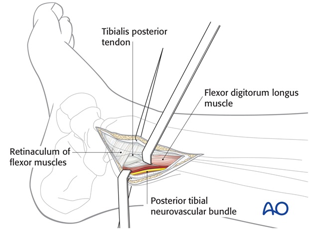
Retract the tendons of the tibialis posterior muscle and the flexor digitorum longus muscle and the posterior tibial neurovascular bundle, using blunt retractors.
Develop the dissection until the fracture line at the posterior part of the tibia is entirely in view.
Dissect the periosteum only as far as required for the control of the reduction at the metaphysis. If necessary, follow the fracture line anteriorly. Protect the saphenous nerve and vein.

5. Pearl
If the fracture line extends very far to the lateral part of the distal tibia, it can be helpful to modify the approach. In this case, carry out dissection between the tendons anteriorly and the neurovascular bundle posteriorly to expose the posterior tibia.


