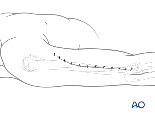
Lateral approach to the humeral shaft
1. Introduction
The lateral approach allows safe exposure of the distal two thirds of the humerus. It can be extended proximally also to expose the proximal humerus.
2. Skin incision
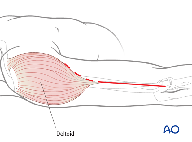
The incision follows a line from the deltoid insertion to the lateral epicondyle.
It may be extended proximally along the anterior, or rarely posterior, margin of the deltoid.
3. Superficial dissection
Elevate the posterior skin and subcutaneous tissue flap away from the deep fascia to ensure that the deep fascia over the triceps can be incised posterior to the lateral intermuscular septum.
4. Deep dissection
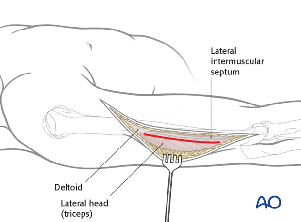
Elevate the anterior flap of the muscular fascia from the edge of the lateral intermuscular septum to prepare for later exposure of the anterior compartment of the arm.
At this stage of the approach it is important to enter the posterior compartment of the arm between the triceps muscle and the lateral intermuscular septum.
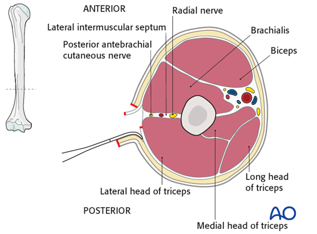
After entering the posterior compartment, find the posterior antebrachial cutaneous nerve, which can be followed proximally to the radial nerve. (Distally this cutaneous nerve passes through the fascia over brachioradialis.)
Develop the interval between the lateral intermuscular septum and triceps, distally to proximally.
In the middle third of the humerus find the radial nerve within fat immediately adjacent to the triceps.
Pearl: As a rough orientation, the radial nerve enters the anterior compartment between the distal and middle third of the humerus.
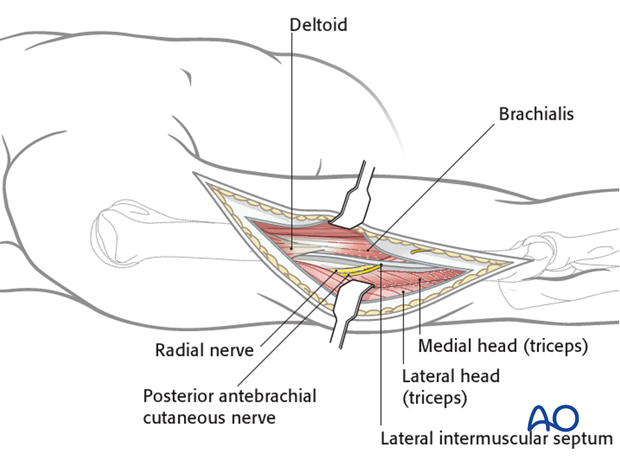
Distally, follow the radial nerve between brachialis and brachioradialis in the anterior compartment of the arm. Carefully retracting the nerve, expose the underlying humerus. Release the lateral intermuscular septum, as needed, for access to the bone and to mobilize the radial nerve.
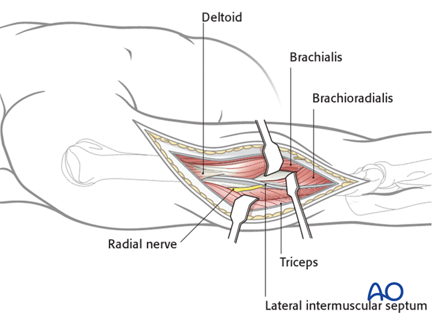
Follow the radial nerve proximally, posterior to the humerus and anterior to the triceps.
This allows safe exposure of the distal two thirds of the humerus.
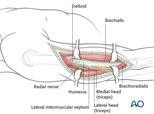
5. Proximal extension of the approach
If necessary, extend the dissection proximally along the deltopectoral interval to access the proximal humerus. This does not allow access to the proximal part of the radial nerve.
Alternatively, extend the incision along the posterior deltoid and into the posterior compartment to follow the radial nerve, but the bone exposure is more limited.
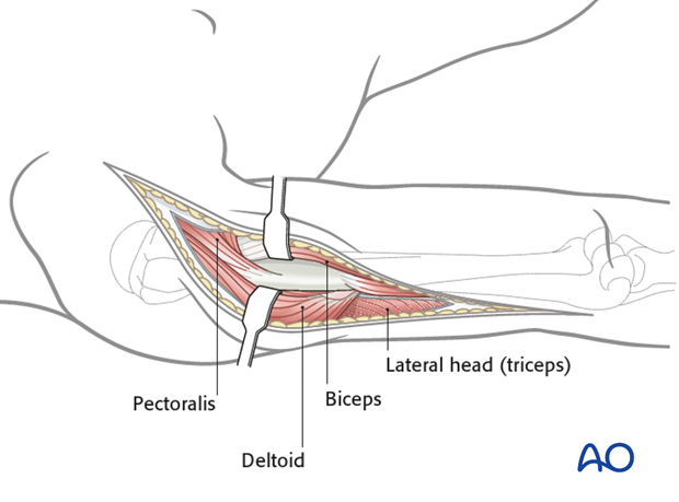
6. Wound closure
Close the incision in layers in a standard manner.
