Highlights of intraoperative imaging
Introduction
The content of the intraoperative imaging compendium is based on the AO Imaging curriculum. For use in AO Surgery Reference, the corresponding course material has been adapted and extended by Daniel Rikli, with Simon Lambert as the editor.
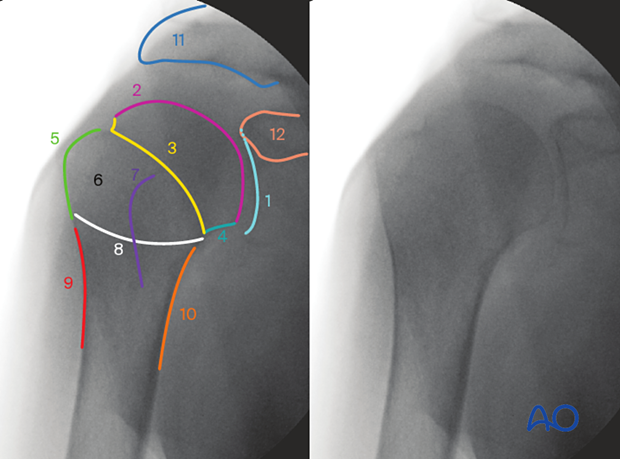
Each joint module – from the shoulder down to the ankle in the adult trauma skeleton – contains now information on how to obtain optimal images. Color marks help to recognize and understand radiographical landmarks.

AO TV 2022: Interview with Daniel Rikli
AO Surgery Reference—Intraoperative Imaging
Daniel Rikli talks with Hannah Shellswell about the revisions to intraoperative imaging in AO Surgery Reference, the history of the AO Imaging Curriculum, what makes intraoperative imaging of a joint so challenging, how to avoid the pitfalls when taking and interpreting a fluoroscopy (focus on the proximal femur) and, how the new content helps to improve the outcome of a surgery.
He outlines how the idea, sparked by a challenging question of a resident, evolved through gathering scientific data to a full-scale course curriculum with the goal of teaching surgeons to improve their work.
AO teaching video
Intraoperative Imaging of the Proximal Femur
Daniel Rikli provides insight on tips and tricks in intraoperative imaging of the proximal femur. (21 minutes)
Imaging curriculum
The AO Imaging curriculum has been developed by the following contributors:
- Proximal humerus: Michael Kraus, Mike Gardner, Maxim Privalov, Sven Vetter, Jochen Franke
- Elbow: Sebastian A Müller, Lars Adolfsson, Nils Beisemann, Antonio Tufi, Daniel Rikli
- Distal radius: Daniel Rikli, Jochen Franke, and Rodrigo Bolaños
- Proximal femur: Daniel Rikli, Michael Blauth, Samir Mehta, and Franz Seibert
- Knee: María Ángela Suárez, Camilo Delgadillo, Julián Salavarrieta, Igor Escalante, Henrik Eckardt, Benedict Swartman, Rodrigo Pesántez
- Ankle: Sven Vetter, Benedict Swartman, Nils Beisemann, and Jochen Franke
Links
On intraoperative imaging, the following learning material is available:
In AO START, there are two learning videos available:


