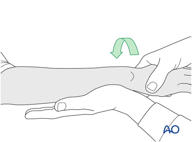
Radial head stabilization (Monteggia fracture-dislocation)
1. General considerations
After anatomical restoration and stable fixation of the ulnar fracture, relocation of the radial head will usually result. If surgery to the radial head is subsequently found to be necessary, it can be approached via a separate incision.
Occasionally, a proximal extension into a Speed and Boyd's approach can be made for additional access to the radial head via a single incision. Care must be taken not to harm the posterior interosseous nerve.
The use of a single approach to both bones of the forearm carries the significant risk of heterotopic bone formation and impairment of forearm function.
Monteggia fracture-dislocation
In Monteggia fracture-dislocations, the ulnar fracture is associated with a dislocation of the radial head.
In most cases, the radial head dislocates anteriorly or laterally; rarely posteriorly.
In Monteggia fracture-dislocations, anatomical reduction and stable fixation of the ulna are mandatory, to ensure stable relocation of the radial head.
Once operative fixation of the ulna has been completed, the surgeon must ensure the stability of the reduced radial head, preferably under image intensification.
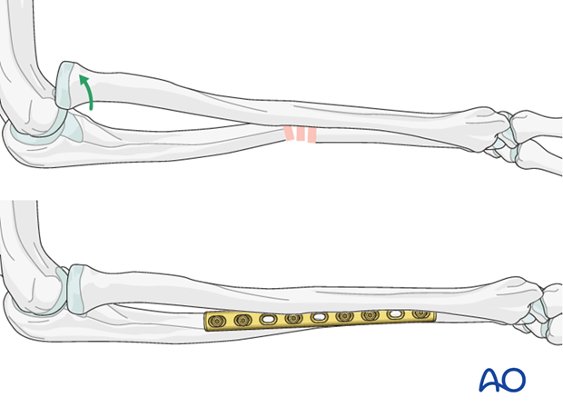
2. Order of reduction and fixation
Anatomic reduction and fixation of the ulna is achieved first, through a standard posterior approach.
Check the position of the radial head, which reduces spontaneously in most of these cases (> 90%). The surgeon must determine the position of forearm rotation in which the radial head is most stable. This is very often in full supination after the radial head has dislocated anterolaterally. The stable rotational position of the forearm is that which will be used when postoperative splintage is applied.
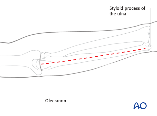
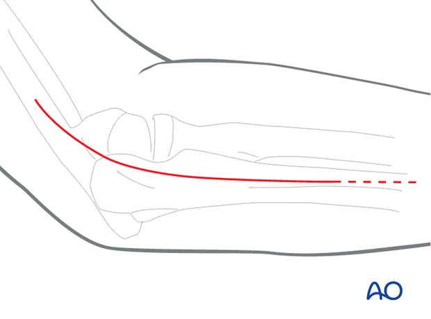
If the radial head does not reduce correctly or if it dislocates on forearm movement (pronation/supination and flexion/extension), this may be due to either malreduction of the ulnar shaft, or there may be interposed soft tissue. If ulnar reduction is confirmed and radial head dislocation persists, access the radial head via a short lateral approach ...
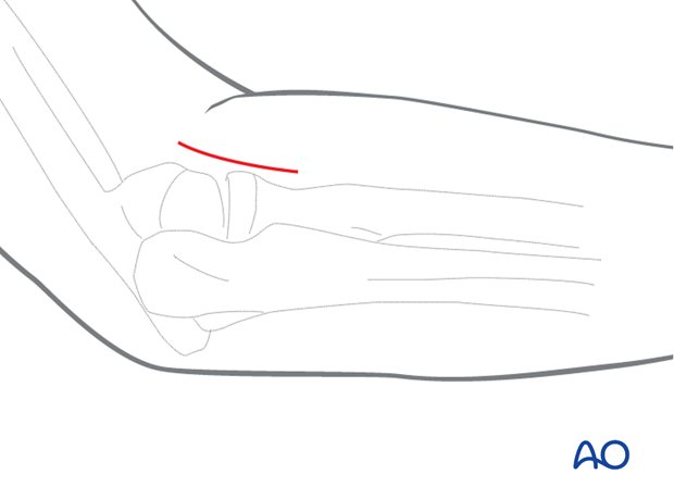
... or an extension into a Speed and Boyd approach (< 10%).
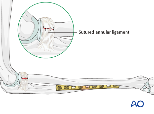
In cases of persisting radial head instability after anatomical fixation of the ulna, interposed annular ligament or the torn joint capsule is usually the cause and should be extracted from the joint and sutured.
3. Monteggia fracture-dislocation
After fixation of the ulna, check the position of the radial head, which reduces in most cases spontaneously. If it did not reduce correctly or if it dislocates on forearm movement (pronation/supination and flexion/extension) …
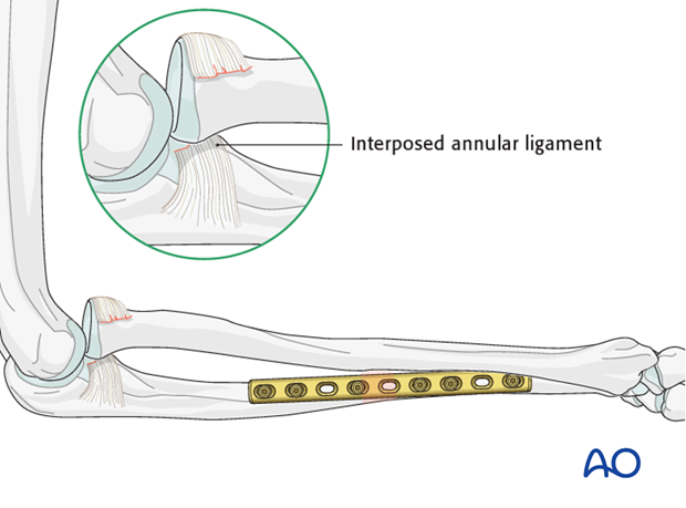
… access the anteriorly dislocated radial head with a lateral approach. In such cases, the usually interposed annular ligament and capsule are extracted and repaired.
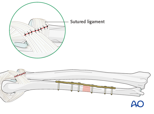
If the head was posteriorly dislocated, a Speed and Boyd’s approach is used to suture the dorsal capsular ligamentous defect.
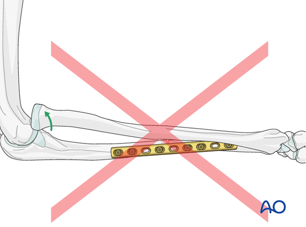
Pitfall: persisting instability of radial head
Be aware that malreduction of the ulna will lead to insufficient spontaneous anatomical reduction and/or instability of the radial head.
Such a situation demands revision of the ulnar fixation if optimal forearm function is to be restored.
4. Check of osteosynthesis
Check the completed osteosynthesis by image intensification. Make sure that the plate is at a proper location, the screws are of appropriate length and a desired reduction was achieved.
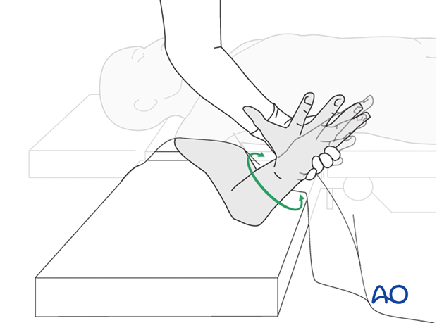
Confirm the rotational position which affords maximal stability of the radial head. This will be the forearm position in which postoperative forearm splinting will be applied.
5. Assessment of Distal Radioulnar Joint (DRUJ)
Before starting the operation the uninjured side should be tested as a reference for the injured side.
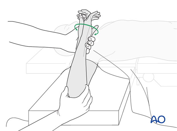
After fixation, the distal radioulnar joint should be assessed for forearm rotation, as well as for stability. The forearm should be rotated completely to make certain there is no anatomical block.
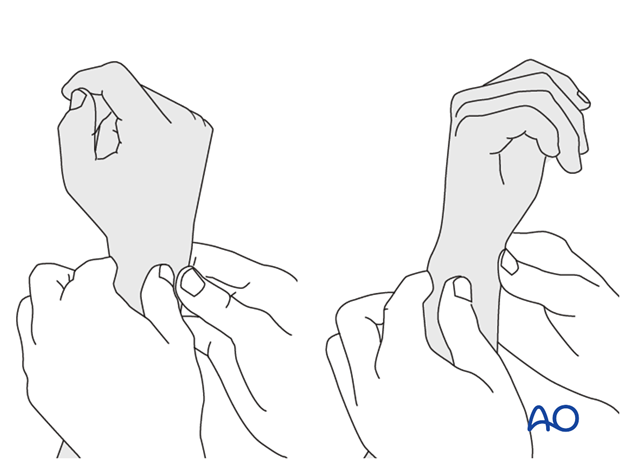
Method 1
The elbow is flexed 90° on the arm table and displacement in dorsal palmar direction is tested in a neutral rotation of the forearm with the wrist in neutral position.
This is repeated with the wrist in radial deviation, which stabilizes the DRUJ, if the ulnar collateral complex (TFCC) is not disrupted.
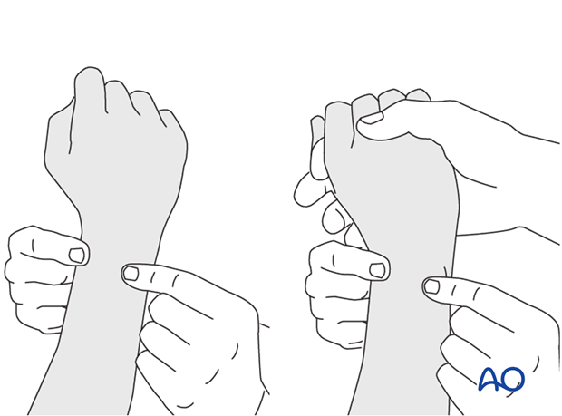
This is repeated with the wrist in full supination and full pronation.
Method 2
In order to test the stability of the distal radioulnar joint, the ulna is compressed against the radius...
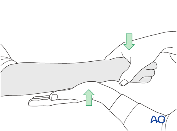
...while the forearm is passively put through full supination...
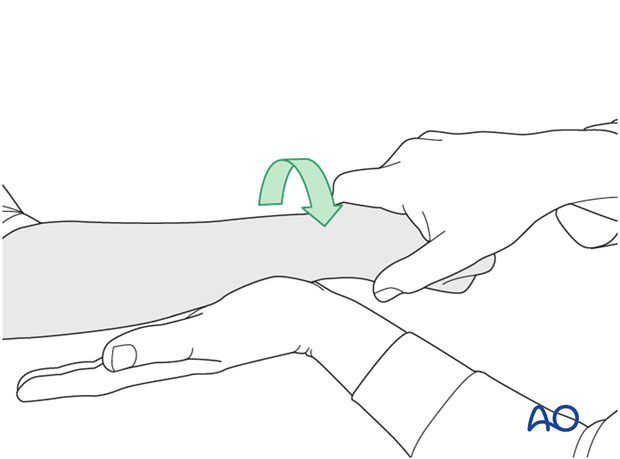
...and pronation.
If there is a palpable “clunk”, then instability of the distal radioulnar joint should be considered. This would be an indication for internal fixation of an ulnar styloid fracture at its base. If the fracture is at the tip of the ulnar styloid consider TFCC stabilization.
