Supine position with knee flexed
1. Preliminary remark
This patient position is used for intramedullary nail fixation of distal tibia fractures.
Intramedullary nail fixation is useful for some distal tibia fractures. Reduction requires careful planning, positioning, and mechanical aids. The fracture must be reduced before the nail is placed across it. A tourniquet is rarely necessary.
2. Radiolucent table
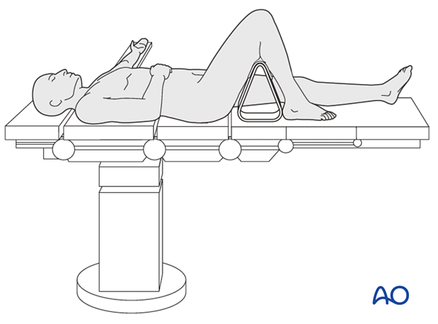
The patient is placed in supine position. The knee of the injured leg is flexed at least 90°. The thigh is supported on a padded rest so that the fracture may be manipulated. Alternatively, an external fixator or distractor may hold the fracture reduced while the foot rests on the table surface. The uninjured leg is extended.
The image intensifier is placed on the opposite side of the table.

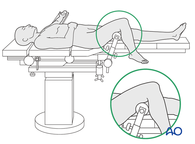
Radiolucent table with knee support
If a radiolucent table with a knee support is used, the padding should be under the distal femur (not compressed under the popliteal fossa).

3. Traction table
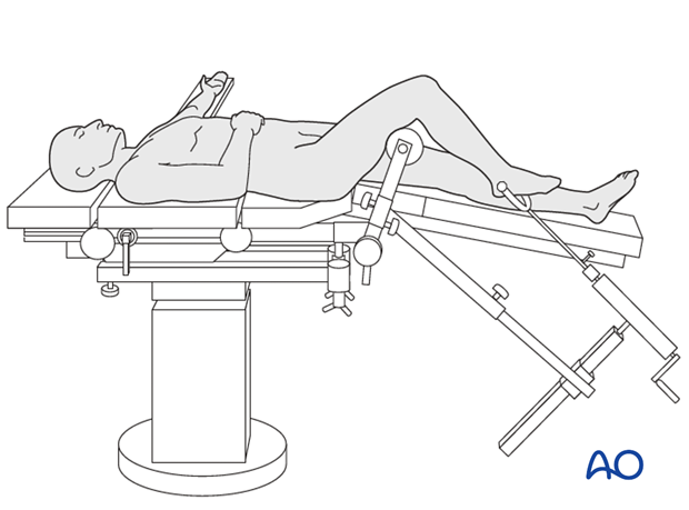
A traction table may be helpful, particularly if a skilled assistant is not available. Countertraction is provided by well-padded support under the distal femur.
The leg position should allow AP and lateral fluoroscopy with the image intensifier on the opposite side of the table.


