Anatomical concepts
1. Bony anatomy
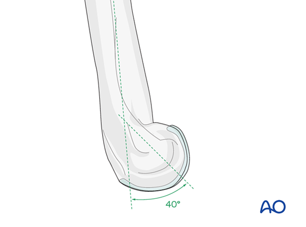
Articular block
The articular block of the distal humerus is anteriorly angled by about 40° with respect to the humeral shaft axis.
Stability of the distal humerus
The articular block and both columns form an asymmetric triangle, which is the base for the stability of the distal humerus.
The columns embrace the articular block from each side.
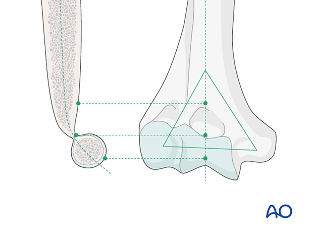
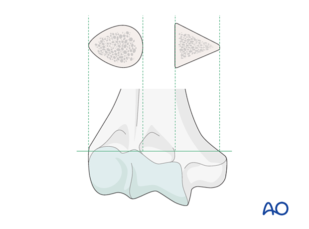
The lateral column has a teardrop cross-section shape (ie, it resists torsion better).
The medial column has a triangular cross-section shape (and therefore is susceptible to shearing and torsion forces).
Distal articular surface
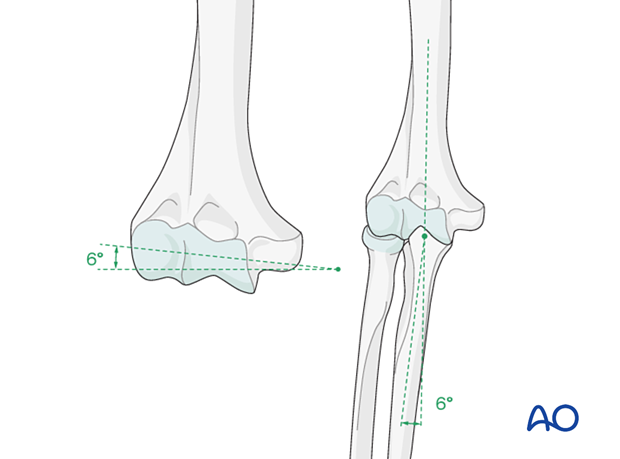
The medial part of the trochlea extends more distally than the lateral part and the capitellum, resulting in a normal valgus “carrying angle” of the elbow joint. With the elbow extended, the long axis of the forearm creates a mean 6° angle to the long axis of the humerus in the coronal plane. This carrying angle is usually greater in females.
Comparison with the intact contralateral elbow is helpful to assess correct angulation.
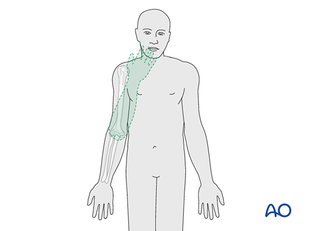
During elbow flexion, the forearm moves on a plane such that the hand goes directly towards the mouth. Any changes in the valgus alignment after the reduction may distort the original plane of movement.
2. Soft-tissue anatomy
Periarticular stabilizing muscles
All muscles that cross the elbow joint impart a compressive joint reaction force.
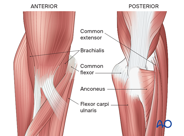
- The common flexor and common extensor muscles contribute to the stability of the elbow under varus and valgus forces, respectively.
- The anconeus muscle, which lies in the same plane as the lateral ulnar collateral ligament, contributes to posterolateral rotatory stability of the ulnohumeral joint.
- The common flexor muscles, on the medial aspect of the elbow, protect the medial ulnar collateral ligament complex from excessive valgus strain and help to stabilize the ulna on the trochlea.
- The brachialis and triceps provide sagittal plane stability by their action on the anterior and posterior aspects of the joint.
Extraarticular stabilizing muscles
- The biceps muscle acts primarily to supinate the forearm, acting through the proximal radioulnar joint.
- The triceps has a muscular (medial head) and tendinous (long head) attachment to the olecranon and aponeurotic expansion (lateral head), which is in continuity with the deep fascia covering the anconeus muscle.
Ligaments
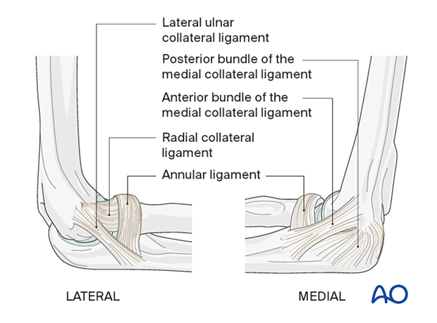
- The collateral ligaments originate from the condyles of the humerus and insert into the ulna. They limit the rotation and varus/valgus movements of the ulna on the trochlea.
- The radius is co-apted to the ulna by the annular ligament, which blends with the lateral ulnar collateral ligament and inserts onto the supinator crest of the ulna.
- The medial capsule has thickenings of which the anterior band of the medial collateral ligament is the most important resisting valgus displacement in extension of the elbow.
- The posterior bundle of the medial collateral ligament resists internal rotatory forces on the ulnohumeral joint.
- After fracture fixation, rehabilitation is principally aimed at preventing stiffness due to capsular contraction.
