Posterior triceps-elevating approach (after Bryan and Morrey) to the distal humerus
1. Introduction
The triceps-elevating approach offers an extensile posterior exposure to the elbow joint by reflecting the triceps insertion from the olecranon.
This is an approach primarily used for arthroplasty.
A disadvantage of this approach is the need to repair and protect the triceps at the end of the procedure.
Consequently, there is a risk for triceps insufficiency.
2. Skin incision
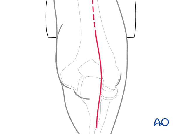
Make an incision centered on the junction of the middle and distal thirds of the humeral shaft. Avoid placing the incision over the tip of the olecranon. Some surgeons make a straight incision slightly medial or lateral, whereas others prefer a curved incision. The incision ends over the ulnar diaphysis.
Elevate full-thickness fasciocutaneous flaps to protect the cutaneous nerves.
3. Ulnar nerve mobilization
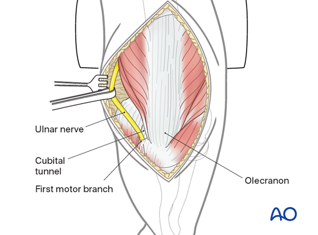
Identify the ulnar nerve proximally along the medial border of the triceps.
Release the ulnar nerve through the cubital tunnel up until the first motor branch by incising the flexor-pronator aponeurosis as the nerve passes between the two heads of flexor carpi ulnaris.
Whenever possible, take care to preserve the perineural vessels.
The nerve may be transposed or left in situ according to the surgeon’s preference, but it should be tension free and not in contact with suture material or metalwork at the end of the procedure.
Take care to protect and be mindful of the nerve throughout the entire procedure.
4. Extensor apparatus
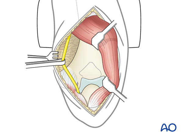
Detach the triceps insertion subperiosteally from the proximal ulna towards the radial side.
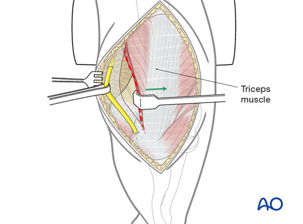
Incise the posterior capsule proximal to the olecranon.
At the level of the olecranon, detach the extensor apparatus subperiosteally or with a sliver of bone using a fine osteotome.
Release the extensor muscles from the lateral epicondyle of the humerus and the anconeus from the posterolateral humerus and ulna. Now the entire extensor apparatus flap can be retracted to the radial side.
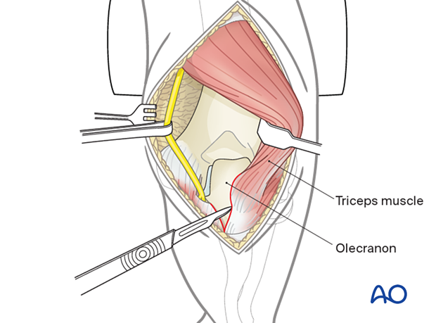
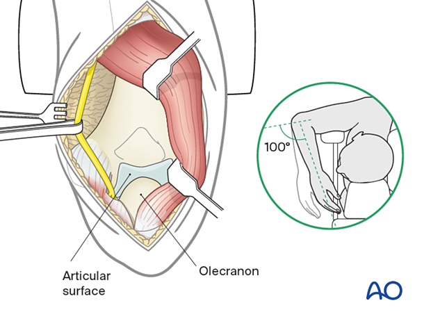
Flex the elbow beyond 100° to enhance visualization of the articular surface.
Optionally, the tip of the olecranon may be removed.
5. Wound closure
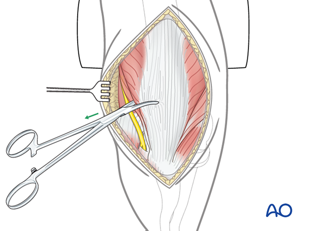
Pull the extensor apparatus into place using a Kocher forceps.
Some surgeons place the ulnar nerve back in the cubital tunnel, whereas others perform an anterior subcutaneous transposition.
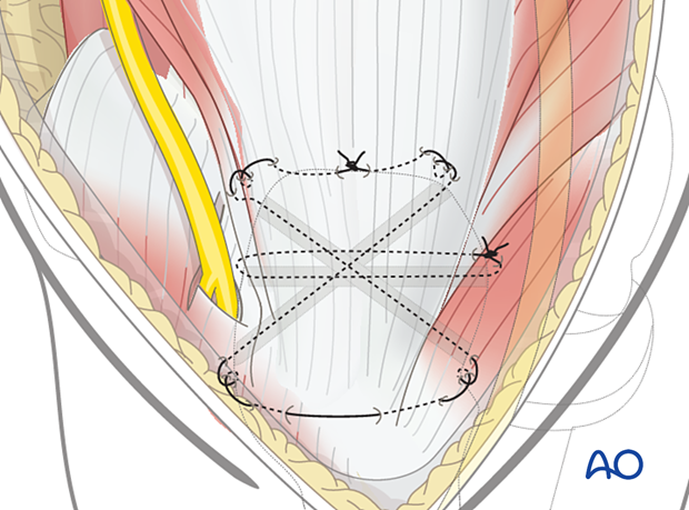
Reattach the triceps tendon insertion to the olecranon with transosseous sutures.
Start by drilling the transosseous tunnels in the olecranon using a 2–2.5 mm drill bit.
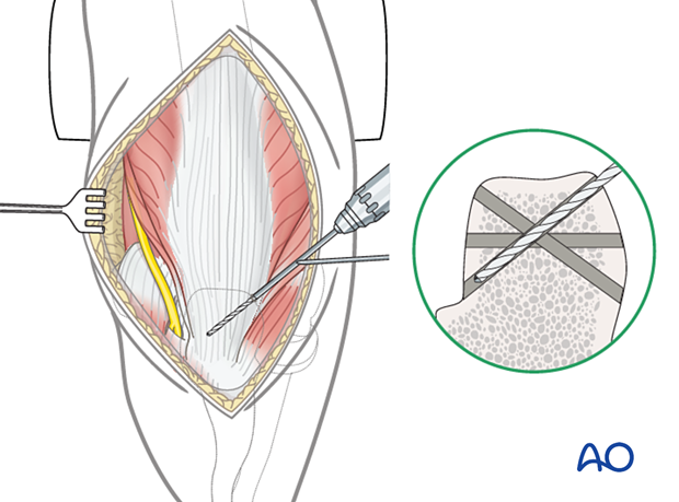
Place heavy nonabsorbable braided sutures through these tunnels.
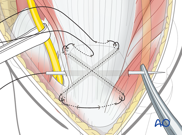
Secure the triceps tendon and fascia with locking sutures and surgical knots. This provides compression of the triceps tendon to the olecranon.
The patient should avoid resisted extension activities while the tendon heals to avoid triceps insufficiency.
