Anterior approach to the capitellum to the distal humerus
1. Introduction
The anterior approach can be used to access fractures of the capitellum, although it is more common to use a laterally based approach for this type of fracture.
2. Skin incision
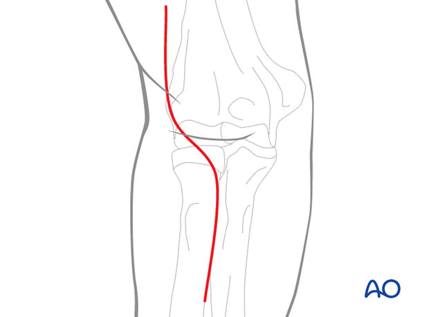
Perform a curved incision over the anterior aspect of the elbow. Proximally this lies along the lateral edge of the biceps muscle belly, and distally lies along the medial border of the brachioradialis of the forearm.

3. Superficial dissection
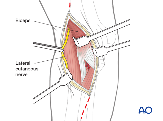
Incise the fascia over the biceps muscle belly and retract the biceps medially.
Identify and protect the lateral cutaneous nerve of the forearm.

4. Deep dissection
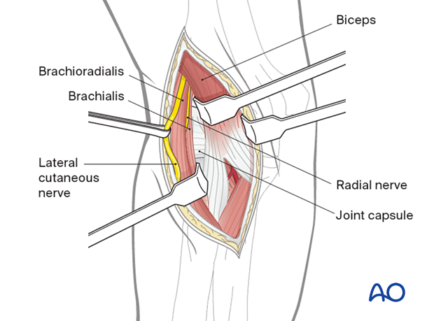
Identify the interval between brachioradialis and brachialis. Within this interval, the radial nerve should be identified and traced distally until it branches into the posterior interosseous branch (PIN) and the superficial radial nerve.
The brachioradialis and brachialis can now be safely retracted with protection of the radial nerve to allow identification of the anterior joint capsule.

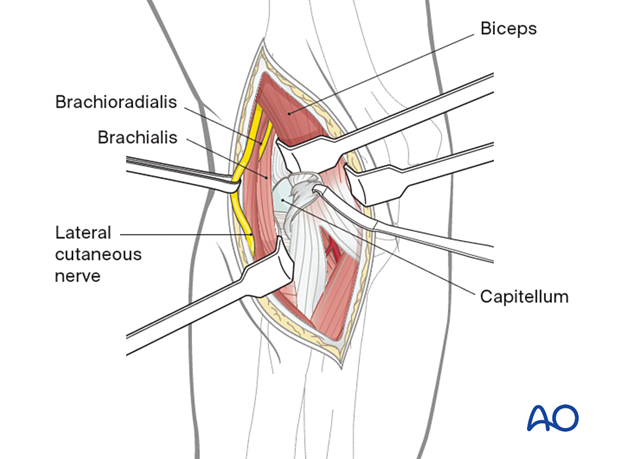
Incise the joint capsule longitudinally to allow access to the capitellum and lateral trochlear ridge.

5. Wound closure
Close the wound in layers.


