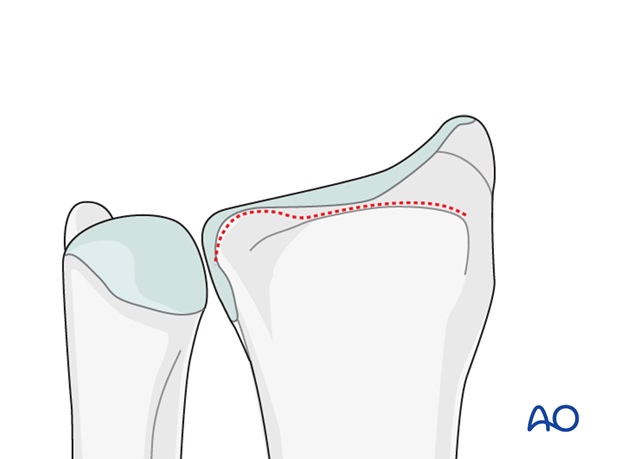
Anatomy of the distal forearm
A thorough knowledge of the anatomy around the wrist is essential. The following images give a short introduction.
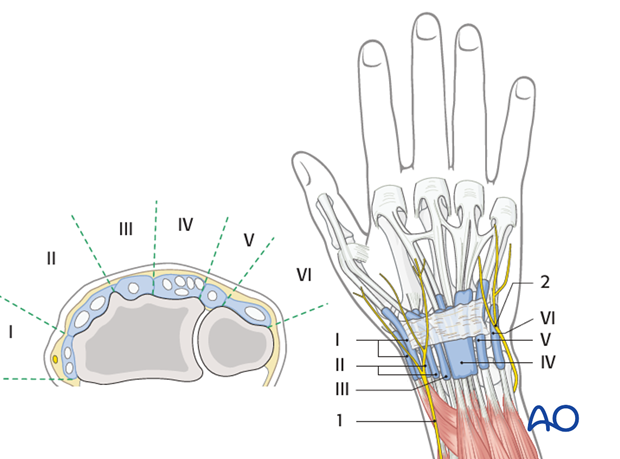
1. Soft tissue anatomy - dorsal
Extensor compartments:
I. Abductor pollicis longus and extensor pollicis brevis
II. Extensor carpi radialis longus and brevis
III. Extensor pollicis longus
IV. Extensor digitorum communis and extensor indicis proprius
V. Extensor digiti minimi
VI. Extensor carpi ulnaris
Nerves:
- Superficial radial nerve
- Dorsal branch of ulnar nerve
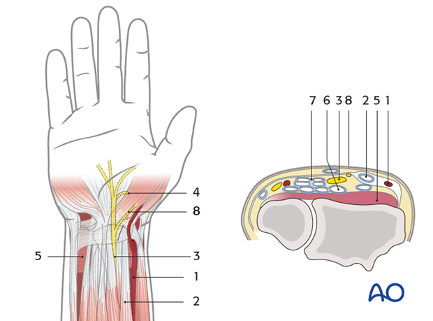
2. Soft tissue anatomy - palmar
- Radial artery
- Flexor carpi radialis tendon
- Median nerve
- Motor branch of the median nerve
- Pronator quadratus muscle
- Flexor digitorum profundus tendons
- Flexor digitorum superficialis tendons
- Palmar cutaneous branch of the median nerve
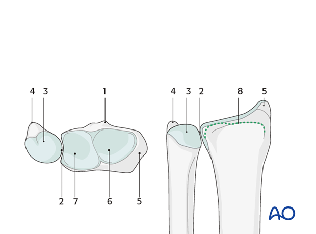
3. Bony anatomy
- Lister's tubercle
- Sigmoid notch
- Ulnar head
- Ulnar styloid
- Radial styloid
- Scaphoid facet
- Lunate facet
- Watershed line
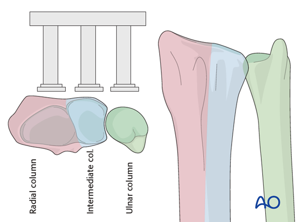
4. Principle of columns
The distal forearm may be thought of in terms of three columns. The ulna forms one column. The radius may be thought of as an intermediate and a radial column.
Distally at the wrist joint, the radial column articulates with the scaphoid and the intermediate column articulates with the lunate.
The ulnar column terminates distally at the TFCC.
Watershed line
The watershed line represents the margin between the structures which may be elevated proximally and the capsule of the wrist joint which should be respected.
