Medial approach to the distal femur
1. Principles
The medial approach to the distal femur is useful to expose medial distal femoral fractures, a Hoffa-type fracture.
It is also useful to expose the neurovascular bundle when a distal femoral fracture is complicated by an arterial injury.
The approach can be extended to expose the posterior cruciate ligament.
This approach also allows limited access to the posterior aspect of the distal femur.
Teaching video
2. Skin incision
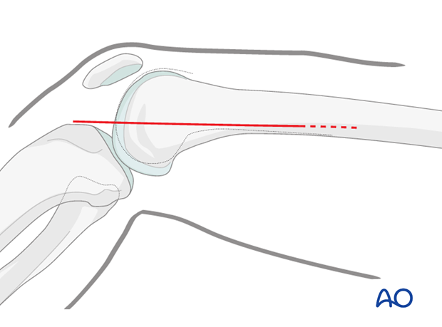
A skin incision is made in the line of the tendon of adductor magnus. The adductor tubercle is identified, and the line of the adductor tendon is marked proximally. A straight-line incision is made along the posterior border of the adductor magnus tendon. The incision can be extended as far proximally as needed.
3. Deep dissection
Identify the anterior edge of the sartorius muscle.
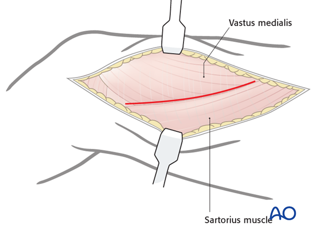
In order to aid the dissection, flex the knee, in order to allow the anterior border of the sartorius to be retracted posteriorly. This will allow the exposure of the tendon of the adductor magnus. The adductor magnus tendon inserts into the adductor tubercle anteriorly.
4. Exposure
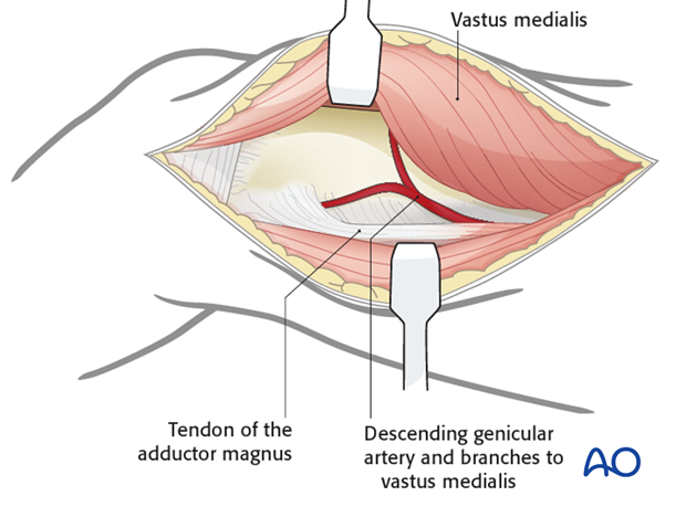
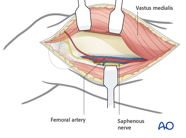
Retract adductor magnus muscle and tendon posteriorly and retract the vastus medialis anteriorly to expose the femur.
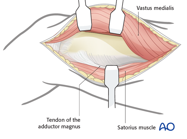
The popliteal neurovascular bundle lies in the popliteal space behind the femur and, if necessary, can be exposed through this approach. Blunt dissection behind adductor magnus facilitates this. A capsulotomy can be made if inspection of the joint surface is needed, but it can be difficult to get a satisfactory access to the posterior joint surface.
5. Wound closure
After careful hemostasis, the wound is closed in layers with absorbable sutures and the chosen skin closure.


