Clinical and radiological examination
1. General considerations
Any signs and symptoms involving the shoulder region require an additional focused evaluation to assess distal pulses, motor and sensation, as well as active and passive shoulder motion where possible. Any potential associated injuries of the entire shoulder girdle and upper extremity should be considered followed by a focused clinical examination of the clavicle and shoulder.
Fractures of the medial end of the clavicle
Recognition of significantly displaced fractures, especially in the posterior or retrosternal direction requires urgent assessment and treatment due to potential injuries to the underlying neurovascular structures and airway. Medial end fractures are often missed. These fractures are rare and may present as stress fractures from repetitive strain/activity in pathologic or osteoporotic bone, or from high energy blunt/penetrating trauma with multisystem injuries. These patients may have increased mortality due to the severity of their other life-threatening injuries.
2. Clinical examination
Clinical assessment starts with inspection of the clavicle and shoulder. The skin is assessed for any tenting or open wounds.
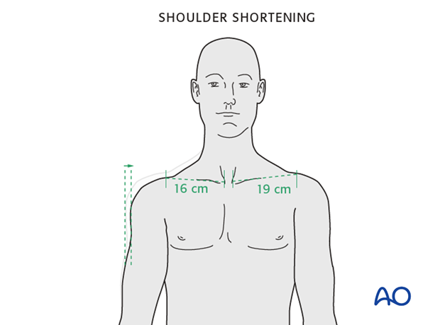
Shortening of the clavicle can be identified by comparison to the opposite intact side by comparing the distance between two fixed bony landmarks such as the medial end of the clavicle (which is easily palpable) and the acromioclavicular joint.
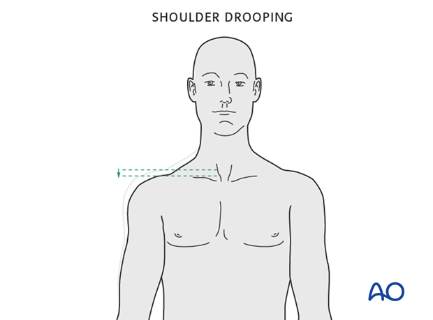
Asymmetric shoulder drooping may also be evident.
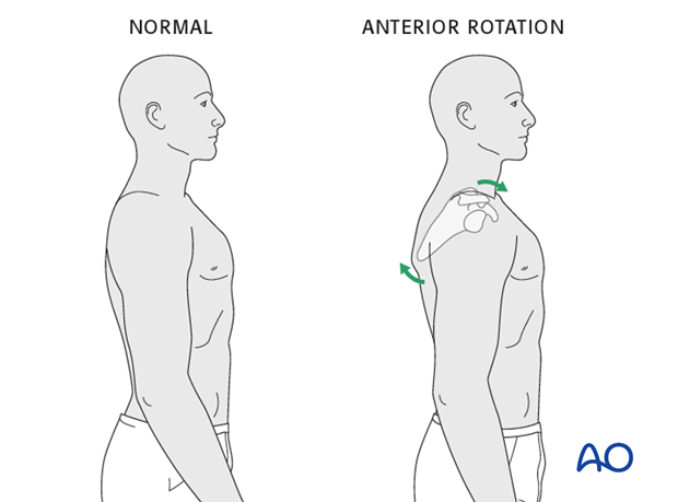
Anterior rotational deformity may be indicated by the presence of ptosis of the shoulder and/or scapular winging.
3. Radiographic assessment
After the patient’s clinical status has been established and stabilized, x-ray examination of the injured shoulder is mandatory. AP X-rays of the clavicle may underestimate the degree of injury or displacement.
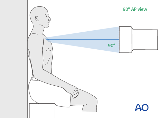
To access radiographically the entire clavicle, specific views are required depending on the location of the injury.
AP view
- Medial
- Shaft
- Lateral
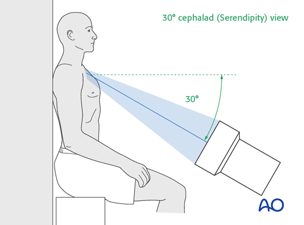
30° cephalad (Serendipity) view
- Medial
- Shaft
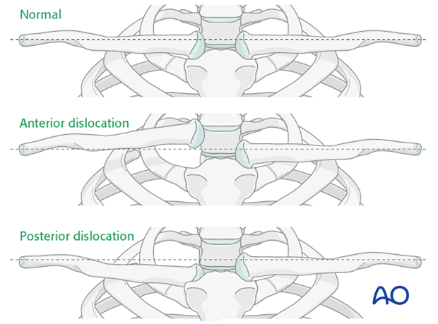
This illustration shows the typical sternoclavicular dislocation patterns seen in a serendipity projection.
For medial end clavicle injuries, CT imaging is often obtained to access better the fracture in the axial plane i.e. posterior/retrosternal displacement which is difficult to access on plain radiographs.


