ORIF - Screw fixation
1. Diagnosis
General considerations
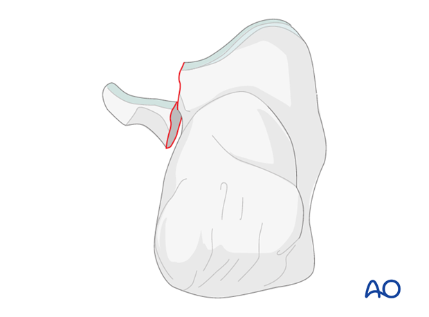
This fracture occurs with a fall from height, usually accompanied by a twisting mechanism. Sometimes this injury is minimized or missed and it is not until continued symptoms push for further investigation that a diagnosis is made.
Radiology
X-rays
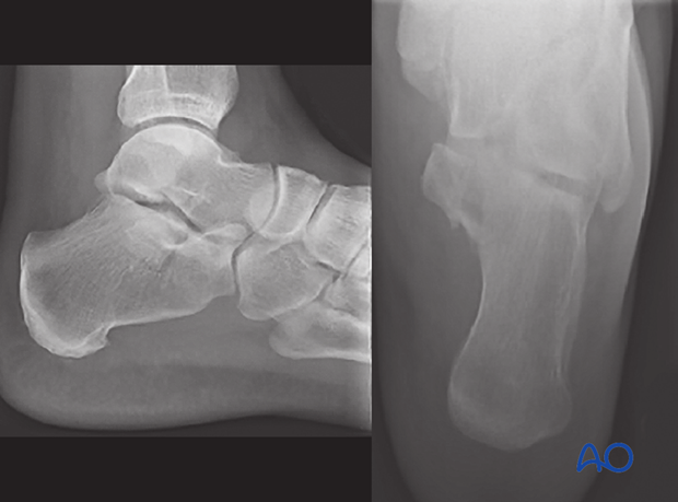
The key to this injury’s diagnosis involves careful and correct x-ray and x-ray interpretation. Lateral x-rays of the hindfoot and axial views are sufficient, but often misinterpreted, or minimized.
CT
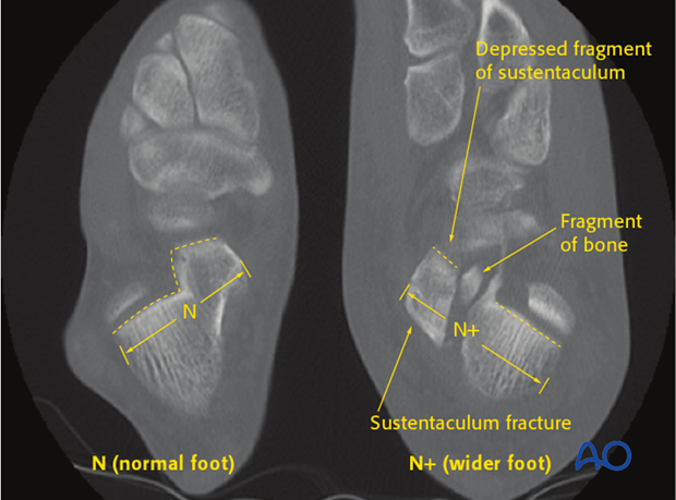
Initially, the diagnosis of this fracture is usually missed because it is difficult to pick up on normal x-rays of the foot. Continuing pain often leads to a CT scan. The diagnosis then becomes obvious.
As this injury occurs with axial load, the sustentaculum remains attached to the talus and is depressed plantarwards immediately with the os calcis and the foot migrating laterally and upwards.
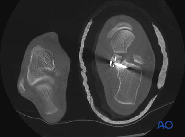
As seen with the normal CT on the left, the middle facet should always be more superior and not depressed, as is demonstrated by the CT on the right. Note the tremendous widening of the hindfoot.
2. Indications for surgery
As this injury makes the three facets of the subtalar joint incongruent, most sustentacular fractures, when displaced, require surgery. Only undisplaced fractures and fractures in the very elderly would be treated nonoperatively. Accurate reduction is almost always indicated for good subtalar function.

3. Approach
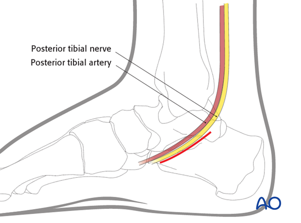
For this procedure a medial approach to the calcaneus may be used.
4. Reduction and fixation
This fracture is plantarly displaced. Like all displaced joint injuries, it requires buttress plating and/or lag screw fixation. Minifragment fixation is appropriate.
Usually, a good surgical approach in which the fracture morphology is recognized, direct reduction of the sustentaculum, provisional K-wire fixation and careful assessment of the reduction by means of image intensification will lead to a good result.
Reduction: tips & tricks
This fracture represents a very small piece of bone, but a very important joint surface. As in most depressed fractures, it will have a cortical surface that needs to be reduced in order to establish correct height. Once height is maintained with temporary K-wire fixation, definitive buttress plating and lag-screw fixation can be utilized. The reduction is not easy because the fragment is displaced inferiorly and has to be elevated sufficiently to be anatomically reduced. The elevation is often very difficult because one pushes upwards on the sustentaculum instead of pulling downwards and medially on the os calcis. This maneuver is complex, but in order to obtain an anatomic reduction, the normal relationship has to be restored. The middle facet must always be more proximal when reduced than the posterior facet, as seen on an axial CT (see diagnosis).
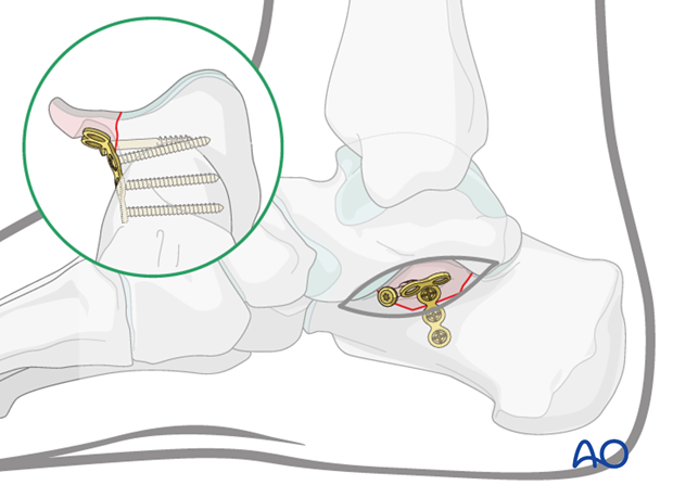
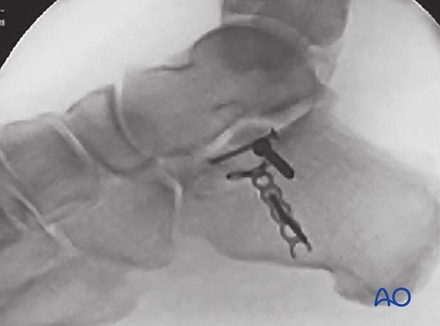
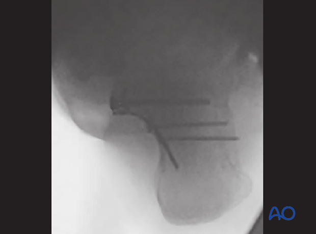
Immediate postop images
Postoperative CT
Note on this postoperative CT that normal relationship of the middle facet has been restored. It is now more proximal than the posterior facet. The medial plate is buttressing the sustentacular fragment which was first reduced and fixed with a lag screw. The compression provided absolute stability. The medial plate in the buttress position prevents axial displacement.
5. Aftertreatment
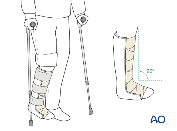
Initially, the patient’s foot and ankle are kept in a well-padded posterior splint that maintains the foot in a neutral position. Surgical drains are removed 2 days after surgery.
The leg should be elevated intermittently through the first number of weeks. The ankle joint is put through range-of-motion exercises as soon as possible, usually by 2-5 days.
Weight bearing is delayed until there is radiographic evidence of union.
Radiography including lateral and axial views is obtained at 6,12 and 26 weeks.
Teaching video
AO teaching video: Application of a lower leg backslab splint


