Intraoperative imaging
1. Introduction
Indications
Whenever an intraoperative CT scanner is available, an intraoperative scan should be obtained for intraoperative evaluation of the reduction.
Any zygomatic fracture benefits from intraoperative CT scanning to visualize fracture reduction and to determine the need for orbital wall reconstruction after zygoma reduction.
When using computer assisted surgery, the reduction (and fixation if necessary) is performed according to standard procedures as described in the AO Surgery Reference. It should be considered an adjunct to surgical treatment.
Computer assisted surgery in treating fractures allows intraoperative visualization of the reduction using intraoperative imaging combined with image fusion of preoperative and intraoperative CT scans.
In simple orbital floor fractures radiopaque material for orbital wall reconstruction can easily be visualized by intraoperative imaging allowing intraoperative or postoperative verification of proper reconstruction.
With this technique, insufficient fracture reduction can be can be identified and corrected, eliminating the need for secondary procedures which may be necessary if only postoperative imaging is performed.
Intraoperative imaging requires an additional 10-15 min.
Useful additional reading
2. Preoperative imaging
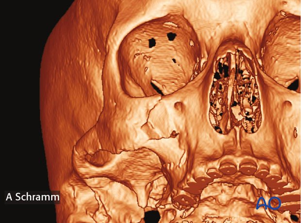
The preoperative CT scan shows a dislocated fracture of the zygomatic complex.

3. Intraoperative assessment of reduction
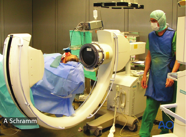
To verify that the zygomatic arch has been properly reduced, a CT scan is performed intraoperatively.

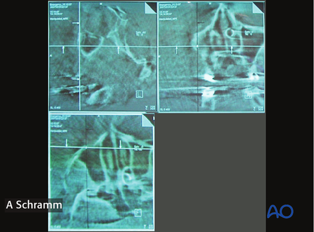
The intraoperative CT scan shows anatomic reduction of the zygomatic complex.
Additionally, correct anatomic shape of the orbital walls can be verified, confirming that there is no need for orbital wall reconstruction.


